

Evaluation of endothelial/Descemet membrane complex of eye bank donor corneas using enhanced depth imaging optical coherence tomography
- PMID: 31190724
- PMCID: PMC6514128
- DOI: 10.2147/OPTH.S185455
Evaluation of endothelial/Descemet membrane complex of eye bank donor corneas using enhanced depth imaging optical coherence tomography
Abstract
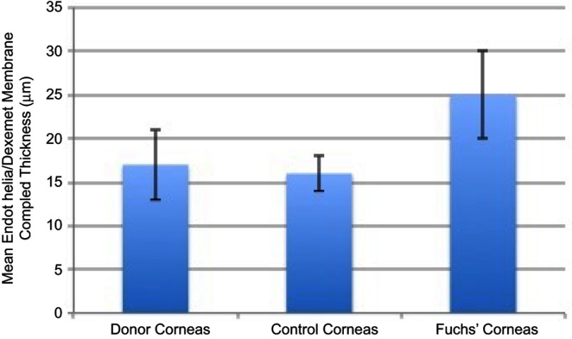
Objective: We present a novel method for screening eye bank donor corneas using high definition optical coherence tomography (HD-OCT). This technology allows for the quantification of endothelial/Descemet membrane (En/DM) complex thickness ex vivo. Design: Prospective interventional study. Participants: Fifty-two corneal grafts from 27 donors were included in this study. Twenty additional control eyes and 11 eyes with Fuchs' endothelial corneal dystrophy were also evaluated for comparison. Methods: A custom built, high speed HD-OCT device (Envisu R2210, Bioptigen, Buffalo Grove, IL, USA) was used to obtain images, and custom-made graph-based segmentation software was used to automatically deconstruct corneal images into micro-layers. HD-OCT imaging was used to scan through the sealed sterile case of donor corneas stored in McCarey-Kaufman medium to image their En/DM complex through the center of the cornea. Results: This technology allowed for quantification of En/DM complex thickness in all donor corneas through the sealed sterile container used to transport graft tissue. Mean En/DM complex thickness of donor corneas was 17±4 μm. The difference between donor cornea En/DM thickness and that of control subjects (16±2 μm) was not statistically significant (p=0.3), suggesting that the transport container and media do not affect measurements. There was a significant difference between En/DM thickness of Fuchs' endothelial corneal dystrophy eyes (25±5 μm) and both donor corneas (p<0.0001) and control subjects (p<0.0001). Conclusions: We have described a new technique to measure En/DM complex thickness in eye bank donor corneas stored in a sealed sterile case. This may represent a novel adjunctive approach to screen corneal grafts for early endothelial disease.
Keywords: Fuchs’ endothelial corneal dystrophy; corneal graft screening; corneal transplant; microscope-integrated OCT.
Conflict of interest statement
United States Non-Provisional Patent (Application No. 14/247903) and United States Provisional Patent (Application No. 62/445,106) (MA). United States Non-Provisional Patents (Application No. 8992023 and 61809518), and PCT/US2018/013409. Patents and PCT are owned by University of Miami and licensed to Resolve Ophthalmics, LLC. MA is an equity holder and sit on the Board of Directors for Resolve Ophthalmics, LLC. Mr Amr Elsawy is listed as a coinventor for a patent 14/247903 licensed to Resolve Ophthalmics. Dr Mohamed Abou Shousha reports grants from National Eye Institute, during the conduct of the study. In addition, Dr Abou Shousha is listed as a coinventor for a patent United States Non-Provisional Patent (Application No. 14/247903) licensed to Resolve Ophthalmics, LLC, a patent United States Provisional Patent (Application No. 62/445,106) (MA). United States Non-Provisional Patents (Application No. 8992023 and 61809518), and PCT/US2018/013409. licensed to Resolve Ophthalmics, LLC. The authors report no other conflicts of interest in this work.
Figures
Similar articles
-
Diagnostic Performance of 3-Dimensional Thickness of the Endothelium-Descemet Complex in Fuchs' Endothelial Cell Corneal Dystrophy.Ophthalmology. 2020 Jul;127(7):874-887. doi: 10.1016/j.ophtha.2020.01.021. Epub 2020 Jan 19. Ophthalmology. 2020. PMID: 32107067
-
Use of ultra-high-resolution optical coherence tomography to detect in vivo characteristics of Descemet's membrane in Fuchs' dystrophy.Ophthalmology. 2010 Jun;117(6):1220-7. doi: 10.1016/j.ophtha.2009.10.027. Epub 2010 Feb 16. Ophthalmology. 2010. PMID: 20163865 Free PMC article.
-
Automated diagnosis and staging of Fuchs' endothelial cell corneal dystrophy using deep learning.Eye Vis (Lond). 2020 Sep 1;7:44. doi: 10.1186/s40662-020-00209-z. eCollection 2020. Eye Vis (Lond). 2020. PMID: 32884962 Free PMC article.
-
Correlation between Guttata Severity and Thickness of Descemet's Membrane and the Central Cornea.Curr Eye Res. 2019 Aug;44(8):849-855. doi: 10.1080/02713683.2019.1600194. Epub 2019 Apr 5. Curr Eye Res. 2019. PMID: 30909752
-
Full-field optical coherence tomography of human donor and pathological corneas.Curr Eye Res. 2015 May;40(5):526-34. doi: 10.3109/02713683.2014.935444. Epub 2014 Sep 24. Curr Eye Res. 2015. PMID: 25251769
Cited by
-
Thickness Measurement of Endothelium-Descemet Membrane in Descemt Membrane Detachment Patients Using High-Definition Optical Coherence Tomography.J Clin Med. 2022 Mar 11;11(6):1534. doi: 10.3390/jcm11061534. J Clin Med. 2022. PMID: 35329859 Free PMC article.
References
Grants and funding
LinkOut - more resources
Full Text Sources
Research Materials
