

The relationship between blood-brain barrier permeability and enlarged perivascular spaces: a cross-sectional study
- PMID: 31190773
- PMCID: PMC6519012
- DOI: 10.2147/CIA.S204269
The relationship between blood-brain barrier permeability and enlarged perivascular spaces: a cross-sectional study
Abstract
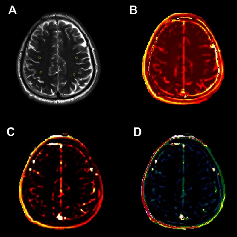
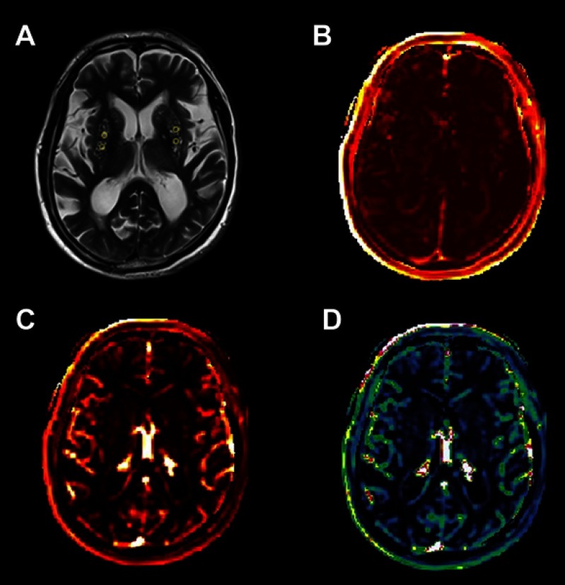
Purpose: Enlarged perivascular spaces (EPVS) have been widely considered as a feature of cerebral small vessel disease (cSVD) but the pathogenesis of EPVS remains unclear. Compromised blood-brain barrier (BBB) integrity may play a role since previous studies have shown that BBB breakdown is a critical contributor to the pathogenesis of other cSVD markers. This study aimed to investigate the association of EPVS in the centrum semiovale (CSO) and basal ganglia (BG) with BBB permeability. Patients and methods: Consecutive participants free of symptomatic stroke history presented for physical examination were enrolled in this cross-sectional study. CSO- and BG-EPVS on T2-weighted (T2-W) magnetic resonance imaging (MRI) were rated using a five-point validated scale. Dynamic contrast-enhanced (DCE)-MRI and Patlak pharmacokinetic model were applied to quantify BBB permeability in the CSO and BG. Results: A total of 109 participants aged 49-90 years (mean age of 69.85 years) were enrolled. The proportions of participants presenting high-grade (>10) EPVS in the CSO and BG were 50.5% and 44.0%, respectively. After adjustments for potential confounders by logistic regression, leakage rate and fractional blood plasma volume were correlated with the severity of BG-EPVS (OR: 5.33; 95%CI: 1.95-14.60 and OR: 0.93; 95%CI: 0.87-0.99). Conclusion: Our study demonstrates that BG-EPVS are associated with compromised BBB integrity, supporting the hypothesis that the BBB dysfunction may be involved in the pathogenesis of BG-EPVS. EPVS in the CSO and BG may have distinct pathophysiology.
Keywords: DCE; MRI; blood–brain barrier; cerebral small vessel disease; dynamic contrast-enhanced; enlarged perivascular spaces; magnetic resonance imaging.
Conflict of interest statement
The authors report no conflicts of interest in this work.
Figures
References
-
- Ding J, Sigurethsson S, Jonsson PV, et al. Large perivascular spaces visible on magnetic resonance imaging, cerebral small vessel disease progression, and risk of dementia: the age, gene/environment Susceptibility-Reykjavik study. JAMA Neurol. 2017. doi: 10.1001/jamaneurol.2017.1397 - DOI - PMC - PubMed
MeSH terms
Substances
LinkOut - more resources
Full Text Sources
