. 2019 May 13:12:3595-3607.
doi: 10.2147/OTT.S184674. eCollection 2019.

miR-106b promotes proliferation and invasion by targeting Capicua through MAPK signaling in renal carcinoma cancer

Affiliations

miR-106b promotes proliferation and invasion by targeting Capicua through MAPK signaling in renal carcinoma cancer

Lu-Jie Miao et al. Onco Targets Ther. .

Abstract

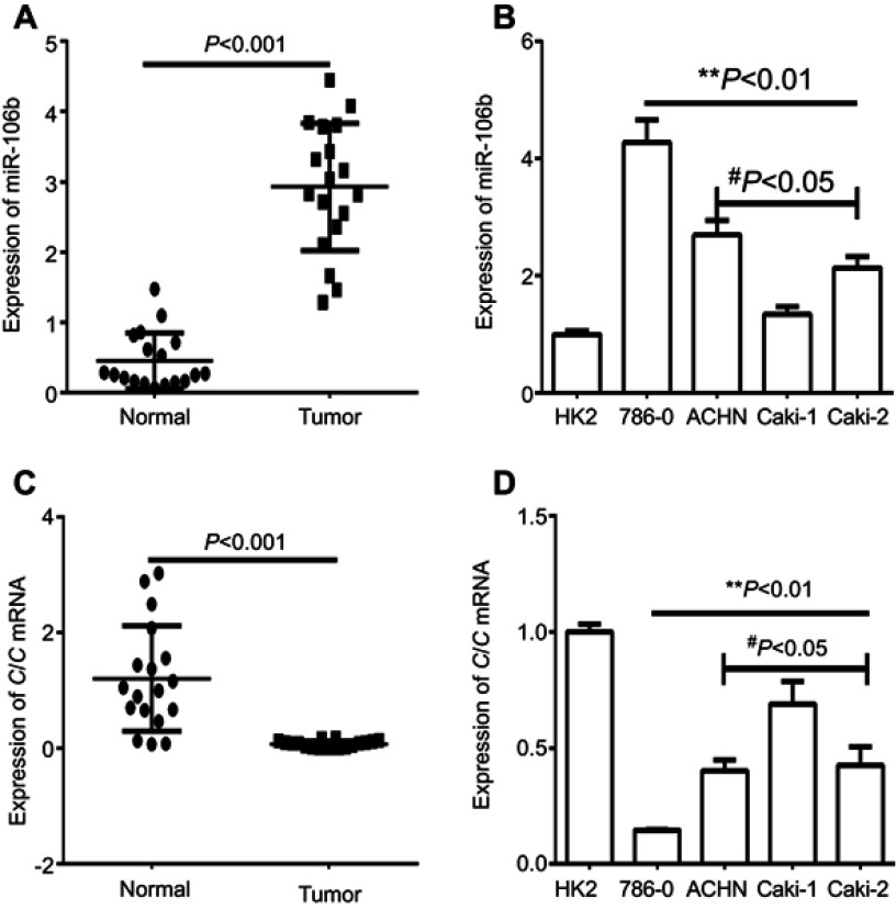
Background: miR-106b has been reported to play a vital role in pathogenesis of some types of cancer, whilst the role of miR-106b in renal carcinoma cancer (RCC) remains unknown. Purpose: The objective of this study was to identify the mechanism of miR-106b regulating the progression of renal carcinoma. Method: The expression of miR-106b was analyzed in RCC cell lines, RCC and adjacent normal renal tissues through qRT-PCR assays. Target mRNA of miR-106b was predicted with databases and verified by luciferase reporter assays. And the effects of miR-106b or targeted mRNA on cell proliferation, invasion, the process of epithelial-mesenchymal transitions (EMTs) were assessed in vitrothrough CCK-8, transwell cell invasion assays, qRT-PCR and Western blotting assays respectively. In addition, the effects of miR-106b on the growth of xenografts mice were analyzedin vivo. Results: The results demonstrated that miR-106b was significantly increased both in RCC tissues and cell lines. Luciferase reporter assays revealed that miR-106b inhibited Capicua expression by targeting its 3'-UTR sequence. And miR-106b promoted cell proliferation, invasion, EMT progression in RCC cellin vitro, as well as promoted the tumor growth of 786-O cells derived xenografts mice. Additionally, loss of Capicua promoted the activation of MAPK signaling pathway. Conclusion: The study suggested that miR-106b regulated RCC progression through MAPK signaling pathway partly by targeting Capicua, which might provide valuable evidence for therapeutic target development of RCC.

Keywords: capicua; epithelial mesenchymal transitions; miR-106b; renal carcinoma.


Conflict of interest statement

The authors report no conflicts of interest in this work.

Figures

Figure 1
Figure 1
Expression of miR-106b and CIC in RCC tissues and cell lines. Notes: (A) qRT-PCR analysis showed the expression of miR-106 in the RCC tissues (tumor) and the adjacent non-tumor tissues (normal) in patient samples (n=20). (B) qRT-PCR analysis of expression of miR-106b in RCC cancer cell lines (786–0, ACHN, Caki-1, Caki-2) and in normal renal tubules epithelium cell HK-2. (C) qRT-PCR analysis showed the expression of CIC in the RCC tissues (tumor) and the adjacent non-tumor tissues (normal) in patient samples (n=20). (D) qRT-PCR analysis of expression of CIC in RCC cancer cell lines (786–0, ACHN, Caki-1, Caki-2) and in normal renal tubules epithelium cell HK-2. **P<0.01, vs HK-2 cell; #P<0.05, vs 786-O cell.Abbreviations: CIC, capicua homology protein; RCC, renal call carvinoma cancer.
Figure 2
Figure 2
Ectopic expression of miR-106b promotes proliferation in RCC cells in vitro and in vivo. Notes: (A) miR-106b levels in 786-O cells transfected with scramble miRNA (miR-NC), miR-106b mimics, or miR-106b inhibitor. (B) CCK-8 assay of 786-O cells transfected with scramble miRNA, miR-106b mimics or miR-106b inhibitor. (C–D) Representative images and quantification of the BrdU cell proliferation assay (400×), *P<0.05, **P<0.01, vs mock group. (E) miR-106b silencing (anti-miR-106b) inhibits tumor growth in 786-O cells derived xenografts compared with negative control (NC). *P<0.05, vs NC.
Figure 3
Figure 3
miR-106b promotes invasion of RCC cells in vitro. Notes: Representative images and quantification of 786-O invasive cells transfected without (Mock) or within scramble miRNA, miR-106b mimics, or miR-106b inhibitor (100×). The cell number of invasive cells were counted in randomly selected fields and presented in the bar graph (mean ± SD; *P<0.05, **P<0.01, vs mock group).
Figure 4
Figure 4
miR-106b promotes EMT progression of RCC cells in vitro. Notes: (A–D) mRNA levels of EMT-related proteins and CIC examined by qRT-PCR in 786-O cells transfected with miR-106b mimics, miR-106b inhibitor, or scramble miRNA. (EI) protein levels of EMT-related proteins and CIC examined by qRT-PCR in 786-O cells transfected with miR-106b mimics, miR-106b inhibitor or scramble miRNA. **P<0.01, vs mock group. Abbreviations: CIC, capicua hohology protein; EMT, epithelial-mesenchymal transition; NC, negative control.
Figure 5
Figure 5
miR-106b directly targets the 3ʹ-UTR of CIC in renal carcinoma cancer. Notes: Luciferase activity of 786-O cells transfected with plasmids carrying a wild-type or mutant 3ʹ-UTR of CIC, in response to miR-106b mimics or miR-NC. **P<0.01, vs miR-NC group. Abbreviations: CIC, capicua homology protein; NC, negative control.
Figure 6
Figure 6
CIC inhibits cell proliferation, invasion in RCC cells. Notes: (A) mRNA levels of CIC in 786-O cells transfected with CIC silencing plasmid (CIC-shRNA), CIC overexpressed plasmid (pcDNA3.1-CIC) or negative controls (pcDNA3.1). (B) CCK-8 assay of 786-O cells transfected with indicated molecular. (C) Representative images and quantification of the BrdU cell proliferation assay (400×). (D) Representative images and quantification of 786-O invasive cells transfected with indicated molecular. *P<0.05, vs mock group; **P<0.01, vs mock group. Abbreviations: CIC, capicua homology protein; RCC, renal cell carcinoma cancer.
Figure 7
Figure 7
CIC inhibits activation of MAPK signaling pathway in vitro. Notes: Changes of phosphorylation levels of MAPK in 786-O cells transfected with indicated molecular. **P<0.01, vs mock group. Representative images (A)  and quantification (B) of  levels of CIC, phosphorylated MAPK and total MAPK  in 786-O cells transfected with indicated molecular.  The  band density was qualified by Image J software, and β-actin was used as internal controls. Abbreviation: CIC, capicua homology protein.

Similar articles

Cited by

References

    1. Chen W, Zheng R, Baade PD, et al. Cancer statistics in China, 2015. CA Cancer J Clin. 2016;66(2):115–132. doi:10.3322/caac.21338 - DOI - PubMed
    1. Siska PJ, Beckermann KE, Rathmell WK, Haake SM. Strategies to overcome therapeutic resistance in renal cell carcinoma. Urol Oncol. 2017;35(3):102–110. doi:10.1016/j.urolonc.2016.12.002 - DOI - PMC - PubMed
    1. Wu YY, Liu XY, Zhuo DX, Huang HB, Zhang FB, Liao SF. Decreased expression of TRPV1 in renal cell carcinoma: association with tumor Fuhrman grades and histopathological subtypes. Cancer Manag Res. 2018;10:1647–1655. doi:10.2147/CMAR.S166390 - DOI - PMC - PubMed
    1. Lee HJ, Shin DH, Kim SY, et al. TFE3 translocation and protein expression in renal cell carcinoma are correlated with poor prognosis. Histopathology. 2018. doi:10.1111/his.13700 - DOI - PubMed
    1. Chen D, Chen W, Xu Y, et al. Upregulated immune checkpoint HHLA2 in clear cell renal cell carcinoma: a novel prognostic biomarker and potential therapeutic target. J Med Genet. 2019;56(1):43–49. doi:10.1136/jmedgenet-2018-105454. - PubMed