. 2019 May 17:12:3881-3891.
doi: 10.2147/OTT.S193433. eCollection 2019.

Mitochondria P-glycoprotein confers paclitaxel resistance on ovarian cancer cells

Affiliations

Mitochondria P-glycoprotein confers paclitaxel resistance on ovarian cancer cells

Weina Guo et al. Onco Targets Ther. .

Abstract

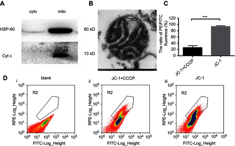
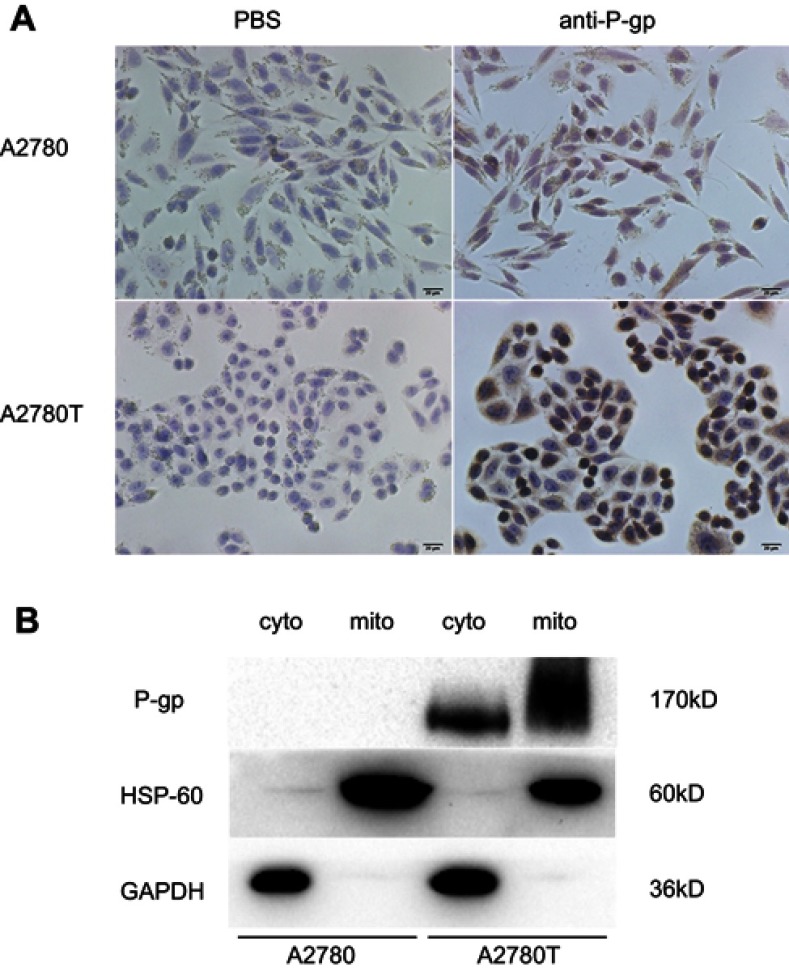
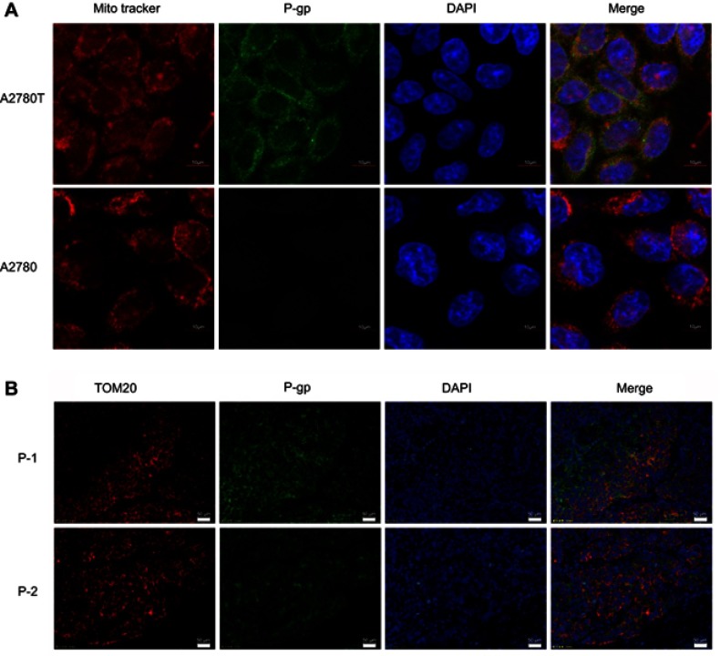
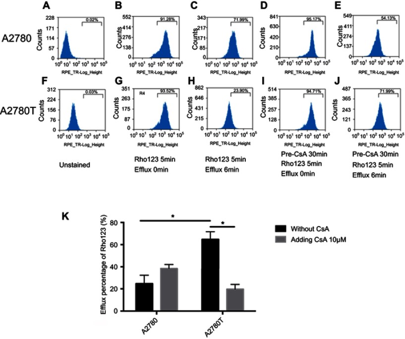
Background: Subcellular expression of P-glycoprotein (P-gp) may play an essential role in multidrug resistance (MDR) in many cancers. However, the mitochondria expression and functional activity of P-gp in ovarian cancer are still unclear. In this study, we isolated mitochondria from A2780 cell line and its paclitaxel-resistant subline A2780T and investigated the expression and function of mitochondria P-gp. Methods: Immunocytochemistry was used to evaluate P-gp expression and subcellular localization in cancer cells. Immunofluorescence and laser confocal microscopy were used to detect the co-localization of P-gp and mitochondria both in ovarian cancer tissues and in cell lines. Western blotting (WB), transmission electron microscopy and JC-1 kit were used to evaluate the purity, integrity and activity of the isolated mitochondria. P-gp expression in the whole cell and the isolated mitochondria was evaluated by WB. Flow cytometry was used to evaluate the efflux function of mitochondria P-gp. Results: P-gp expression was detected at the membrane, cytoplasm and nuclei of the A2780T cells, but not in the A2780 cells. Co-localization of P-gp and mitochondria was observed in the A2780T cell line and ovarian cancer tissues, but not in A2780 cells. The purity, integration and activity of the isolated mitochondria are high. P-gp was highly expressed in the A2780T cells and the isolated mitochondria, but was not found in A2780 cells. Rho123 efflux rate was significantly increased in isolated A2780T mitochondria compared to those in A2780 (43.2% vs 9.6%), but it was partly reversed by cyclosporin A (CsA, a P-gp inhibitor). Conclusion: P-gp is highly expressed in mitochondria of taxol-resistant ovarian cancer cells and ovarian cancer tissues and mediates the drug efflux, which probably protect cancer cells from chemotherapy.

Keywords: P-glycoprotein; mitochondria; multidrug resistance; ovarian carcinoma.


Conflict of interest statement

The authors report no conflicts of interest in this work.

Figures

Figure 1
Figure 1
Identification the purity, integrity and activity of isolated mitochondria. Notes: (A) Western blot analysis of HSP-60 (1:1000) and Cyt c (1:1000) in cytosolic and mitochondria fractions of A2780 and A2780T cell lines. (B) the sediment of isolated mitochondria was processed with fixation, dehydration, embedding, section. Electron microscopic image showed the integrated mitochondria isolated from A2780 cell line. Bar, 200nm. (C) isolated mitochondria were processed with PBS, JC-1+CCCP (10uM) and JC-1 respectively for 15min at 37℃. Mitochondria with pre-added 10uM CCCP (ii) had significantly low ratio of PE/FITC (21.2%) compared with the mitochondria without adding CCCP (iii) (94.6%) (P<0.001). ***P<0.001. The images are representative of at least three independent experiments with similar results.
Figure 2
Figure 2
The expression of P-gp in A2780 and A2780T cells. Notes: (A) immunocytochemistry staining of P-gp in A2780 cell and A2780T cells. P-gp was expressed in plasma membrane, cytosol, and nuclei in A2780T cells, but not in A2780 cells. (B) Western blot analysis of P-gp in cytosolic and mitochondrial fractions of A2780 and A2780T cell lines. HSP-60 and GAPDH were used as a protein loading control of mitochondria and cytosol, respectively. Three separate experiments were done.
Figure 3
Figure 3
Co-localization of P-gp and mitochondria in A2780T cell line and clinical biopsies by confocal laser scanning and fluorescence microscopy. Notes: (A) Red fluorescence showed the localization of mitochondria and green fluorescence showed the expression of P-gp were overlaid to form fluorescent orange, which represents the co-localization of P-gp and mitochondria. Bar, 10μm. (B) Nuclei were dyed in blue fluorescence. P-1: patient 1 is a patient who relapsed within six months after the end of six cycles of chemotherapy treatment. P-2: patient 2 is a patient who had no recurrence or progression within six months after the end of six cycles of chemotherapy treatment. Bar, 50μm. TOM20 is the maker protein located in mitochondrial outer membrane.
Figure 4
Figure 4
Flow cytometry assays show the active efflux mediated by mitochondrial P-gp. Notes: After being incubated with the Rho123, the mitochondria isolated from A2780 and A2780T with or without pre-treatment with CsA were compared to the efflux of Rho123. In the unstained mitochondria, PBS was used instead of Rho123 (A, F). According to whether adding P-gp inhibitor CsA or not, the isolated mitochondria were divided into two groups: control group (B, C, G, H) and intervention group (D, E, I, J). In the control group, the Rho123 intake rate of mitochondria isolated from both cell lines increasing beyond 90% (B, G). After 6 min efflux, the retained fluorescence of mitochondria isolated from A2780T was significantly lower than that from A2780 (P=0.03; C,H,K). In the intervention group, the retained fluorescence intensity of mitochondria isolated from A2780T was significantly higher than that of the control group (P=0.015; I, J, K). This phenomenon was not found in mitochondria isolated from A2780 (P>0.05; D,E,K). *P<0.05. Three separate experiments were done. Statistical analysis was shown in K. Abbreviation: CsA, cyclosporin A.

Similar articles

Cited by

References

    1. Vaughan S, Coward JI, Bast RC Jr., et al. Rethinking ovarian cancer: recommendations for improving outcomes. Nat Rev Cancer. 2011;11(10):719–725. doi:10.1038/nrc3144 - DOI - PMC - PubMed
    1. Holmes D. Ovarian cancer: beyond resistance. Nature. 2015;527(7579):S217. doi:10.1038/nature15724 - DOI - PubMed
    1. Coleman MP, Forman D, Bryant H, et al. Cancer survival in Australia, Canada, Denmark, Norway, Sweden, and the UK, 1995-2007 (the international cancer benchmarking partnership): an analysis of population-based cancer registry data. Lancet (London, England). 2011;377(9760):127–138. doi:10.1016/S0140-6736(10)62231-3 - DOI - PMC - PubMed
    1. Kunjachan S, Rychlik B, Storm G, Kiessling F, Lammers T. Multidrug resistance: physiological principles and nanomedical solutions. Adv Drug Deliv Rev. 2013;65(13–14):1852–1865. doi:10.1016/j.addr.2013.09.018 - DOI - PMC - PubMed
    1. Fantappie O, Masini E, Sardi I, et al. The MDR phenotype is associated with the expression of COX-2 and iNOS in a human hepatocellular carcinoma cell line. Hepatology (Baltimore, Md). 2002;35(4):843–852. doi:10.1053/jhep.2002.32469 - DOI - PubMed