. 2019 May 17:12:3893-3903.
doi: 10.2147/OTT.S199601. eCollection 2019.

Curcumin reverses oxaliplatin resistance in human colorectal cancer via regulation of TGF-β/Smad2/3 signaling pathway

Affiliations

Curcumin reverses oxaliplatin resistance in human colorectal cancer via regulation of TGF-β/Smad2/3 signaling pathway

Jiahuan Yin et al. Onco Targets Ther. .

Abstract

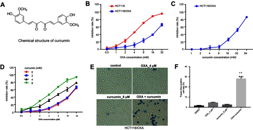
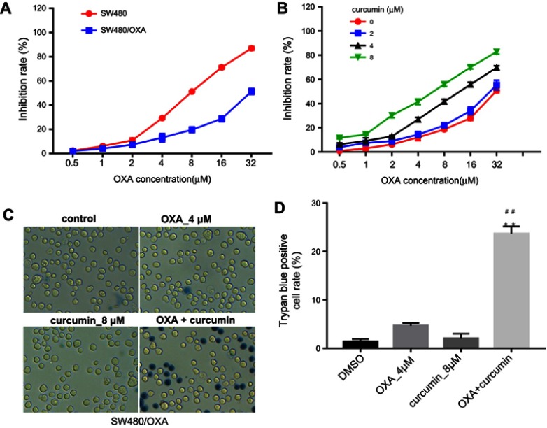
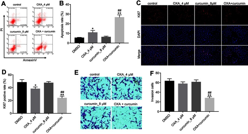
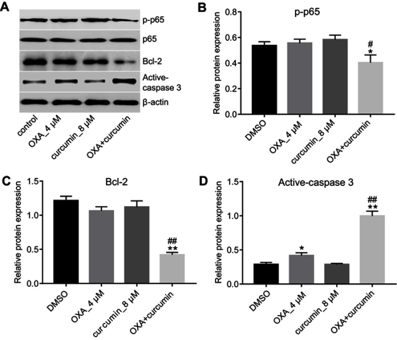
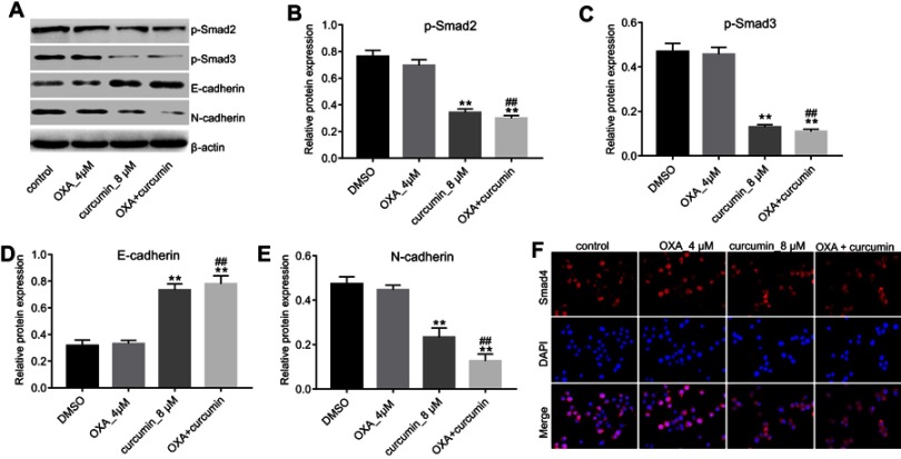
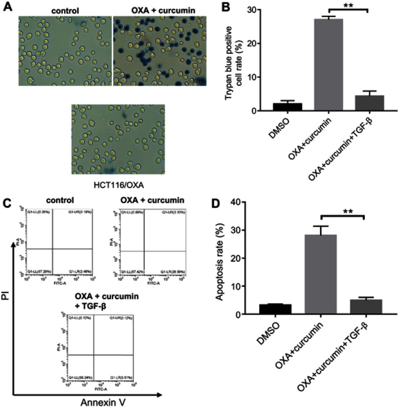
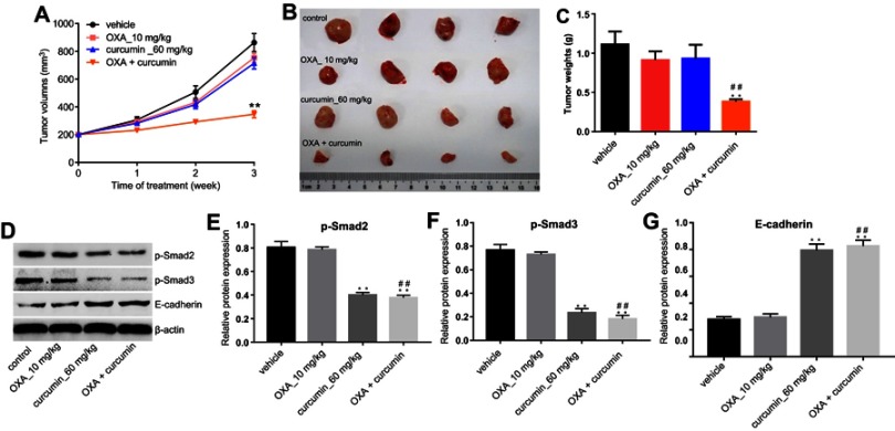
Background: Oxaliplatin (OXA) resistance is a main obstacle to the chemotherapy of colorectal cancer (CRC). Epithelial-mesenchymal transition (EMT), which is mainly regulated by TGF-β/Smad signaling pathway, has gradually been recognized as an important mechanism for tumor chemoresistance. Studies have shown that curcumin regulated EMT processes in many human cancers. However, whether curcumin could regulate OXA resistance in CRC through modulating TGF-β/Smad signaling-mediated EMT remains unclear. Methods: In an attempt to investigate the effect of curcumin on OXA resistance in CRC, OXA-resistant cell line HCT116/OXA was established firstly. The effect of curcumin on cell proliferation was evaluated by MTT assay and Ki67 immunofluorescence staining, respectively. Cell apoptosis was evaluated by flow cytometry. In addition, transwell assay was used to detect the effect of curcumin on cell invasion and the activation of TGF-β/Smad signaling was examined by immunofluorescence and Western blot. Moreover, the therapeutic potential of curcumin was further examined in vivo using a CRC animal model. Results: The OXA-resistant cell line HCT116/OXA was successfully established, and combination of OXA with curcumin reduced OXA resistance in vitro. Besides, the combination treatment inhibited the expressions of p-p65 and Bcl-2, but increased the level of active-caspase3. In addition, curcumin inhibited EMT via regulation of TGF-β/Smad2/3 signaling pathway. Moreover, in vivo study confirmed curcumin could reverse OXA resistance in CRC. Conclusion: Our study indicated that curcumin could reserve OXA resistance in CRC through dampening TGF-β/Smads signaling in vitro and in vivo.

Keywords: TGF-β/Smad signaling pathway; colorectal cancer; curcumin; epithelial-mesenchymal transition; oxaliplatin resistance.


Conflict of interest statement

The authors report no conflicts of interest in this work.

Figures

Figure 1
Figure 1
Establishment of HCT116/OXA cell line and optimization of combination strategy. (A) Chemical structure of curcumin. (B) MTT assay was performed to calculate the growth inhibition rate of HCT116 and HCT116/OXA cells treated with 0.5, 1, 2, 4, 8, 16 or 32 μM OXA for 48 hrs. (C) The growth inhibition rate of HCT116/OXA cells treated with 1, 2, 4, 8, 16, 32 or 64 μM curcumin for 48 hrs. (D) The growth inhibition rate of HCT116/OXA cells treated with the combination of OXA and curcumin. (E) HCT116/OXA cell death was detected with trypan blue staining. (F) Trypan blue positive cell rate in each group was calculated. Each experiment was repeated three times. **P<0.01 compared to control group; ##P<0.01, compared to OXA alone group.
Figure S1
Figure S1
Curcumin reverses oxaliplatin resistance in SW480 cells. (A) MTT assay was performed to calculate the growth inhibition rate of SW480 and SW480/OXA cells treated with 0.5, 1, 2, 4, 8, 16 or 32 μM OXA for 48 hrs. (B) The growth inhibition rate of SW480/OXA cells treated with the combination of OXA and curcumin. (C) SW480/OXA cell death was detected with trypan blue staining. (D) Trypan blue positive cell rate in each group was calculated. Each experiment was repeated three times. **P<0.01 compared to control group; ##P<0.01, compared to OXA alone group.
Figure 2
Figure 2
Curcumin increased OXA-induced growth inhibition in HCT116/OXA in vitro. HCT116/OXA cells were treated with OXA or/and curcumin for 48 hrs. (A) Cell apoptosis was detected with Annexin V/PI staining. (B) Cell apoptosis rate was subsequently calculated with flow cytometry. (C/D) Cell proliferation ability was analyzed by Ki67 immunofluorescence staining assay. (E/F) Cell invasion ability was analyzed by Transwell assay. Each experiment was repeated three times. *P<0.05, **P<0.01 compared to control group; ##P<0.01, compared to OXA alone group.
Figure 3
Figure 3
Curcumin downregulated the expressions of p-p65 and Bcl-2 and upregulated the level of active-caspase3. HCT116/OXA cells were treated with OXA or/and curcumin for 48 hrs. (AD) The relative protein levels of p-p65, p65, Bcl-2 and active-caspase3 in cells were assessed by Western blotting assay. Each experiment was repeated three times. *P<0.05, **P<0.01 compared to control group; #P<0.05, ##P<0.01, compared to OXA alone group.
Figure 4
Figure 4
Curcumin inhibited EMT in CRC through inhibiting TGF-β/Smad2/3 pathway. HCT116/OXA cells were treated with OXA or/and curcumin for 48 hrs. (AE) The relative protein levels of p-Smad2, p-Smad3, E-cadherin and N-cadherin in cells were measured by Western blotting assay. (F) The distribution of Smad4 in cells was detected by immunofluorescence staining assay. Each experiment was repeated three times. **P<0.01 compared to control group; ##P<0.01, compared to OXA alone group.
Figure 5
Figure 5
The inhibitory effect of curcumin plus OXA on the growth of HCT116/OXA cells was completely abolished by TGF-β. HCT116/OXA cells were treated with HCT116/OXA cells were treated with 4 μM OXA plus 8 μM curcumin or 2 ng/mL TGF-β for 48 hrs. (A) HCT116/OXA cell death was detected with trypan blue staining. (B) Trypan blue positive cell rate in each group was calculated. (C) Cell apoptosis was detected with Annexin V/PI staining. (D) Cell apoptosis rate was subsequently calculated with flow cytometry.
Figure 6
Figure 6
Curcumin reduced OXA chemoresistance in CRC by inhibiting TGF-β/Smad2/3 pathway in vivo. HCT116/OXA cells were subcutaneously implanted into nude mice. Mice were divided into four groups: vehicle group, OXA alone group, curcumin alone group and OXA plus curcumin group. (A) Tumor volume of xenograft was measured weekly. (B) The mice were sacrificed in 3 weeks, and the tumors were isolated (C) Quantification of tumor weights in each group. (DG) The relative protein levels of p-Smad2, p-Smad3 and E-cadherin in tumor tissues were measured by Western blot. Each experiment was repeated three times. **P<0.01 compared to control group; ##P<0.01, compared to OXA alone group.

Similar articles

Cited by

References

    1. Siegel RL, Miller KD, Jemal A. Cancer statistics, 2018. CA Cancer J Clin. 2018;68(1):7–30. - PubMed
    1. Petrelli F, Coinu A, Ghilardi M, Cabiddu M, Zaniboni A, Barni S. Efficacy of oxaliplatin-based chemotherapy + bevacizumab as first-line treatment for advanced colorectal cancer: a systematic review and pooled analysis of published trials. Am J Clin Oncol. 2015;38(2):227–233. doi:10.1097/COC.0b013e3182a2d7b8 - DOI - PubMed
    1. Kopetz S, Hoff PM, Morris JS, et al. Phase II trial of infusional fluorouracil, irinotecan, and bevacizumab for metastatic colorectal cancer: efficacy and circulating angiogenic biomarkers associated with therapeutic resistance. J Clin Oncol. 2010;28(3):453–459. doi:10.1200/JCO.2009.24.8252 - DOI - PMC - PubMed
    1. Esatbeyoglu T, Huebbe P, Ernst IM, et al. Curcumin – from molecule to biological function. Angew Chem Int Ed Engl. 2012;51(22):5308–5332. doi:10.1002/anie.201107724 - DOI - PubMed
    1. Chen QH. Curcumin-based anti-prostate cancer agents. Anticancer Agents Med Chem. 2015;15(2):138–156. doi:10.2174/1871520615666150116102442 - DOI - PubMed