

Clinical relevance and significance of programmed death-ligand 1 expression, tumor-infiltrating lymphocytes, and p16 status in sinonasal squamous cell carcinoma
- PMID: 31190998
- PMCID: PMC6514258
- DOI: 10.2147/CMAR.S201568
Clinical relevance and significance of programmed death-ligand 1 expression, tumor-infiltrating lymphocytes, and p16 status in sinonasal squamous cell carcinoma
Abstract
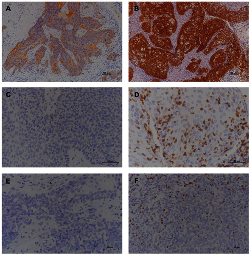
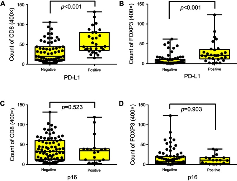
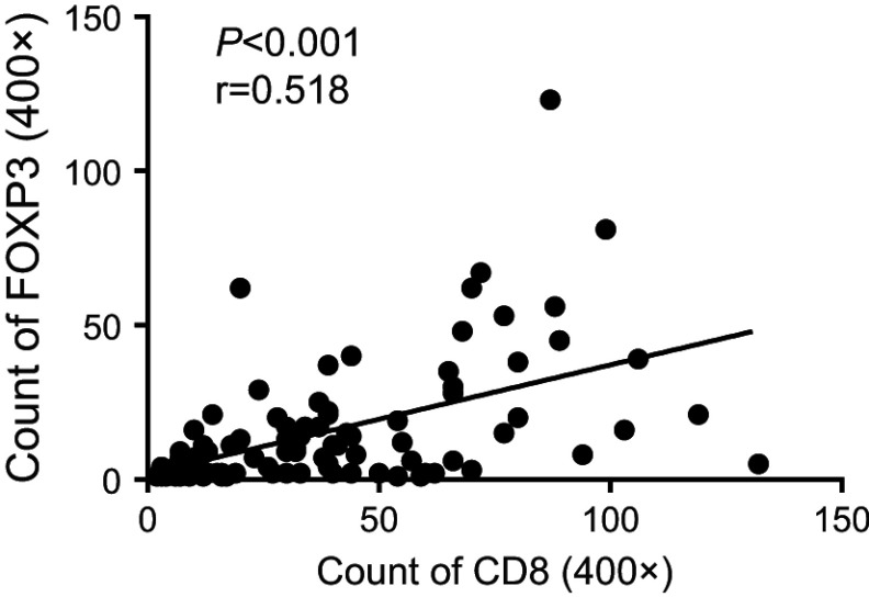
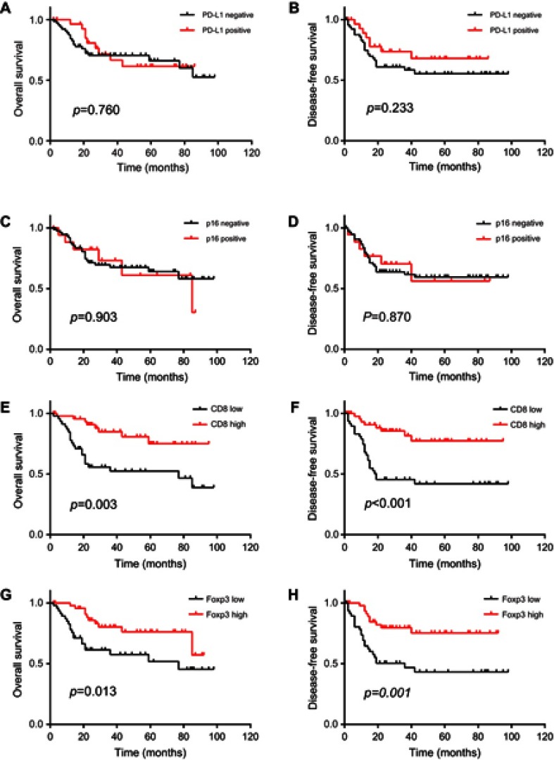
Purpose: Immunotherapy may be a potential alternative for patients with sinonasal squamous cell carcinoma (SNSCC). Data regarding potential immunotherapy targets, such as programmed death-ligand 1 (PD-L1) and tumor-infiltrating lymphocytes (TILs), in SNSCC are limited. In this study, we assessed the prevalence and prognostic value of PD-L1 expression and TILs in p16-negative and p16-positive SNSCC. Patients and methods: Tissues from 96 patients with SNSCC were stained using immunohistochemistry against PD-L1, CD8, and Foxp3 to assess the immune environment. The correlations between PD-L1 expression, TILs, and p16 status were analyzed. Additionally, PD-L1, CD8, and Foxp3 expressions, as well as p16 status, were analyzed in relation to patient clinicopathological variables and prognosis. Results: Twenty-nine (30.2%) patients with SNSCC showed PD-L1 expression in >5% of tumor cells. PD-L1 expression was significantly correlated with poor differentiation and a high level of TILs. PD-L1 expression and the CD8+ and Foxp3+ T-cell infiltrates in p16-negative patients (n=78, 81.2%) and p16-positive patients (n=18, 18.8%) were not significantly different. PD-L1 expression and p16 status were not associated with overall survival (OS) and disease-free survival (DFS). Patients with high CD8+ or Foxp3+ cell infiltration had better clinical outcomes. A multivariate analysis confirmed that CD8 TILs were a significant independent and favorable prognostic factor for OS (p=0.023) and DFS (p=0.008). Conclusion: TILs can play a prognostic role in SNSCC. We did not find differences in immune marker expression between p16-positive and p16-negative SNSCC tissues. The high correlation between PD-L1 expression and TILs indicates that the PD-1/PD-L1 pathway is a promising immunotherapeutic target for SNSCC.
Keywords: biomarker; immunotherapy; prognosis; sinonasal cancer; squamous cell carcinoma.
Conflict of interest statement
The authors report no conflicts of interest in this work.
Figures
References
LinkOut - more resources
Full Text Sources
Research Materials
