

Expression of PD-L1 in cervical carcinoma and its impact on survival associated with T-cell infiltration and FoxP3 expression
- PMID: 31191020
- PMCID: PMC6529624
- DOI: 10.2147/CMAR.S194597
Expression of PD-L1 in cervical carcinoma and its impact on survival associated with T-cell infiltration and FoxP3 expression
Abstract
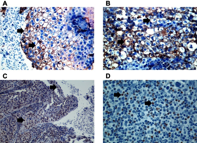
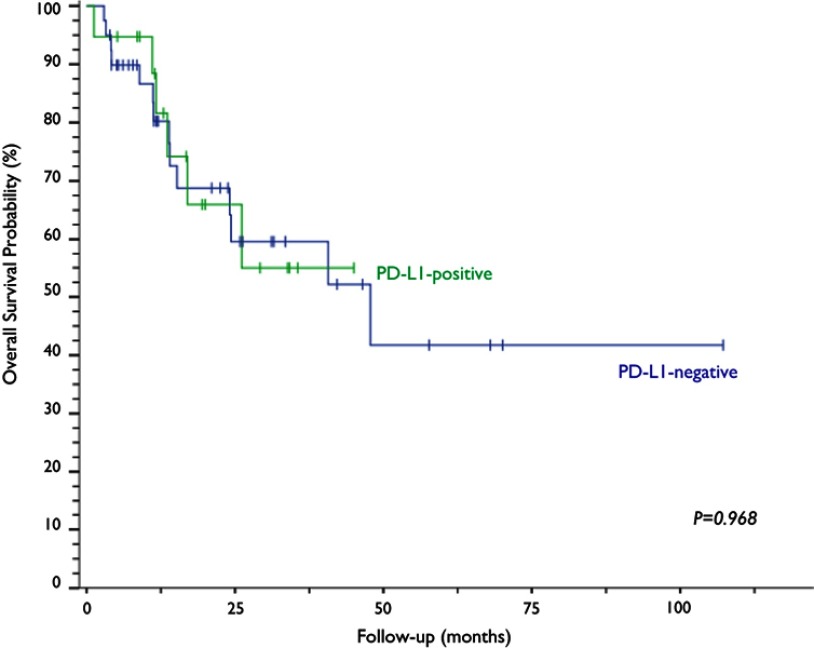
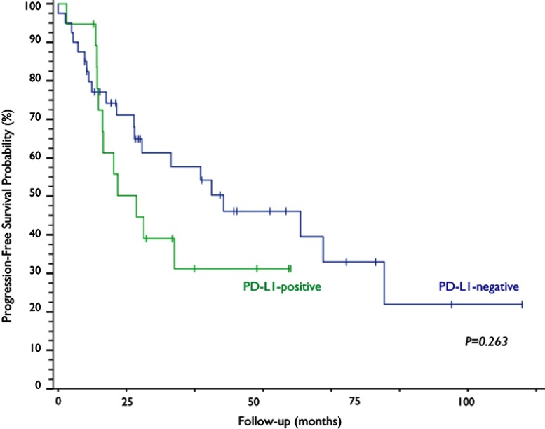
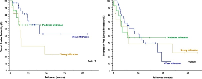
Background: The PD-1/PD-L1 signaling axis is currently the most elucidated mechanism for tumor evasion of T-cell-mediated immunity. Nevertheless, few data are available regarding its impact on cervical cancer and the relationship with lymphocytic infiltrates. Methods: A retrospective assessment of all cases of cervical neoplasia treated in Caxias do Sul General Hospital, Brazil, between 2012 and 2016 was performed. Clinical and pathological data were collected from electronic records and analyzed. Original slides were independently reviewed by three pathologists to confirm diagnoses and to assess the immunohistochemical expression of PD-L1 and FoxP3 in tumor cells and lymphocytic infiltrates. Results: PD-L1 staining was present in 32.2% of the 59 cervical samples. Median overall survival time of the PD-L1-negative group was 47.8 months, a time point not yet reached by the PD-L1-positive group (p=0.968). Median progression-free survival was 24.3 months for PD-L1-negative and 11.5 months for PD-L1-positive patients (p=0.263). PD-L1 staining was found in 27.1% of the lymphocytic infiltrates, and survival analysis revealed no difference between PD-L1-positive and PD-L1-negative samples. There was no impact on survival related to FoxP3 staining in neither tumor samples nor lymphocytic infiltrates. Conclusion: Although the median progression-free survival times differed, the difference was not statistically significant. Our study corroborates the rationale that PD-L1 expression in cervical neoplasms has no impact on survival. PD-L1 expression in peritumoral lymphocytes revealed no impact on infiltration volume nor survival. Keywords: uterine cervical neoplasms, tumor-infiltrating lymphocytes, cancer, tumor microenvironment, survival.
Conflict of interest statement
The authors report no conflicts of interest in this work.
Figures

References
-
- Instituto Nacional De Câncer José Alencar Gomes Da Silva. Estimativa da incidência e mortalidade por câncer no Brasil 2018; 2018. Available from: http://www.inca.gov.br/estimativa/2018/estimativa-2018.pdf. Accessed September 11, 2018.
LinkOut - more resources
Full Text Sources
Research Materials
