Review
. 2019 May 5:2019:1845637.
doi: 10.1155/2019/1845637. eCollection 2019.

Nanoparticle-Based Paramagnetic Contrast Agents for Magnetic Resonance Imaging

Affiliations
Review

Nanoparticle-Based Paramagnetic Contrast Agents for Magnetic Resonance Imaging

Juan Pellico et al. Contrast Media Mol Imaging. .

Abstract

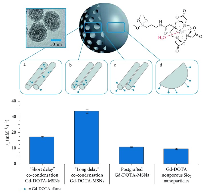
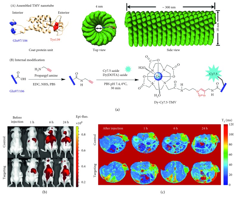
Magnetic resonance imaging (MRI) is a noninvasive medical imaging modality that is routinely used in clinics, providing anatomical information with micron resolution, soft tissue contrast, and deep penetration. Exogenous contrast agents increase image contrast by shortening longitudinal (T 1) and transversal (T 2) relaxation times. Most of the T 1 agents used in clinical MRI are based on paramagnetic lanthanide complexes (largely Gd-based). In moving to translatable formats of reduced toxicity, greater chemical stability, longer circulation times, higher contrast, more controlled functionalisation and additional imaging modalities, considerable effort has been applied to the development of nanoparticles bearing paramagnetic ions. This review summarises the most relevant examples in the synthesis and biomedical applications of paramagnetic nanoparticles as contrast agents for MRI and multimodal imaging. It includes the most recent developments in the field of production of agents with high relaxivities, which are key for effective contrast enhancement, exemplified through clinically relevant examples.


Figures

Figure 1
Figure 1
Typical transmission electron microscope image and schematic representation of Gd-DOTA-MSNs (66.3 ± 6.6 nm) prepared using (a) “short delay” co-condensation, where functionalities are internalised deeply in the structure (r1 = 17.14 ± 0.49 mM−1·s−1), (b) “long delay” co-condensation, where functionalities are internalised nearer to the porous openings (r1 = 33.57 ± 1.29 mM−1·s−1), and (c) postgrafting, where functionalities are loaded on external surfaces (r1 = 10.77 ± 0.22 mM−1·s−1). (d) Postgrafted Gd-DOTA-non-porous silica nanoparticles (r1 = 19.56 ± 0.47 mM−1·s−1) (reproduced from [71]).
Figure 2
Figure 2
Figure illustrating the dual-modal imaging properties of Gd2O3 : Eu3+ nanoparticles (reprinted (adapted) with permission from [48]).
Figure 3
Figure 3
TEM images and size distribution plots obtained from TEM images of (a, b) HoF-el and (c, d) HoF-rh NPs (reprinted (adapted) with permission from [63]).
Figure 4
Figure 4
(a) Schematic illustration for the synthesis of UCNPs-Ce6 and hMUC. (b) In vivo T1-weighted MR images of a tumour-bearing mouse before (left) and after (right) intravenous injection of hMUC. (c) In vivo CT images of a tumour-bearing mouse before (upper) and after (lower) intratumour injection (reprinted (adapted) with permission from [90]).
Figure 5
Figure 5
(a) Structure of the tobacco mosaic virus (TMV) nanoparticle's coat protein with surface-exposed residues highlighted as internal glutamic acid (blue) and external tyrosine (red) and the structure of the assembled capsid/strategy for internal modification. (b) Near-infrared fluorescence (NIRF) imaging of subcutaneous PC-3 (α2β1) prostate tumours in athymic nude mice (n = 3) before and 1, 6, and 24 h after the intravenous injection of Dy-Cy7.5-TMV-mPEG (control group) or Dy-Cy7.5-TMV-DGEA (targeting group). (c) In vivo T2-mapping MRI of subcutaneous PC-3 (α2β1) prostate tumours in athymic nude mice (n = 3) obtained before and 1, 6, and 24 h after the intravenous injection of Dy-Cy7.5-TMV-mPEG (control group) and Dy-Cy7.5-TMV-DGEA (targeting group) (reprinted (adapted) with permission from [62]).

Similar articles

Cited by

References

    1. Frangioni J. V. New technologies for human cancer imaging. Journal of Clinical Oncology. 2008;26(24):4012–4021. doi: 10.1200/jco.2007.14.3065. - DOI - PMC - PubMed
    1. Sanz J., Fayad Z. A. Imaging of atherosclerotic cardiovascular disease. Nature. 2008;451(7181):953–957. doi: 10.1038/nature06803. - DOI - PubMed
    1. Geethanath S., Vaughan J. T., Jr. Accessible magnetic resonance imaging: a review. Journal of Magnetic Resonance Imaging. 2019 doi: 10.1002/jmri.26638. In press. - DOI - PubMed
    1. De León-Rodríguez L. M., Martins A. F., Pinho M. C., Rofsky N. M., Sherry A. D. Basic MR relaxation mechanisms and contrast agent design. Journal of Magnetic Resonance Imaging. 2015;42(3):545–565. doi: 10.1002/jmri.24787. - DOI - PMC - PubMed
    1. Bao Y., Sherwood J. A., Sun Z. Magnetic iron oxide nanoparticles as T1 contrast agents for magnetic resonance imaging. Journal of Materials Chemistry C. 2018;6(6):1280–1290. doi: 10.1039/c7tc05854c. - DOI