. 2019 May 28:13:537.
doi: 10.3389/fnins.2019.00537. eCollection 2019.

The Neuroprotective Effect of Hemin and the Related Mechanism in Sevoflurane Exposed Neonatal Rats

Affiliations

The Neuroprotective Effect of Hemin and the Related Mechanism in Sevoflurane Exposed Neonatal Rats

Fan Yang et al. Front Neurosci. .

Abstract

Background: Many studies have reported that sevoflurane can increase neuronal apoptosis and result in cognitive deficits in rodents. Although neurotoxicity may be associated with mitochondrial dysfunction and oxidative stress, the exact mechanism remains unclear. In order to evaluate potential treatment therapies, we studied the effects of hemin on neurotoxicity of neonatal rat sevoflurane exposure.

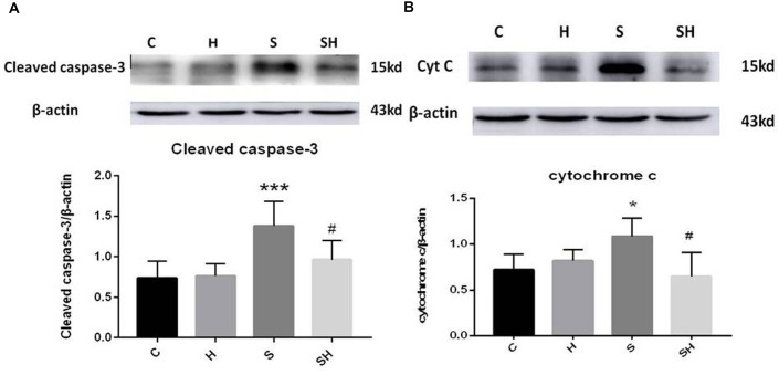
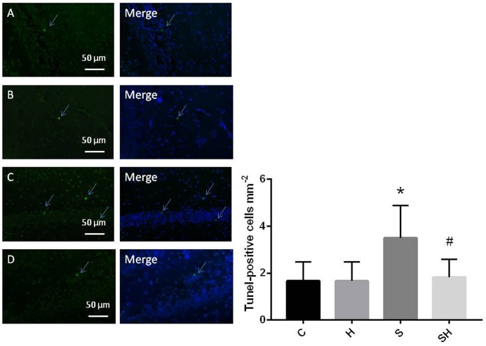
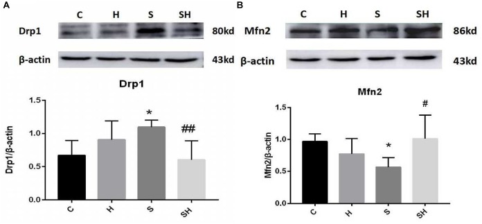
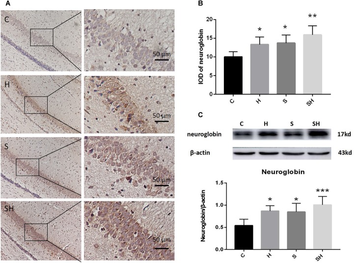
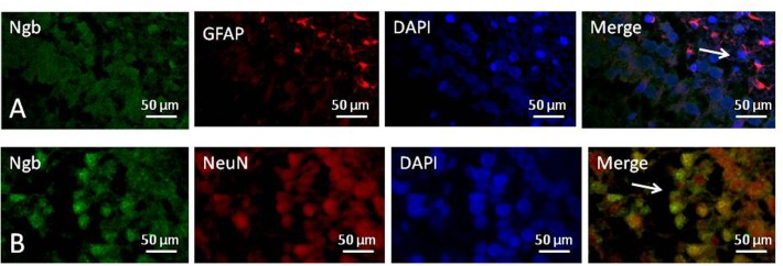
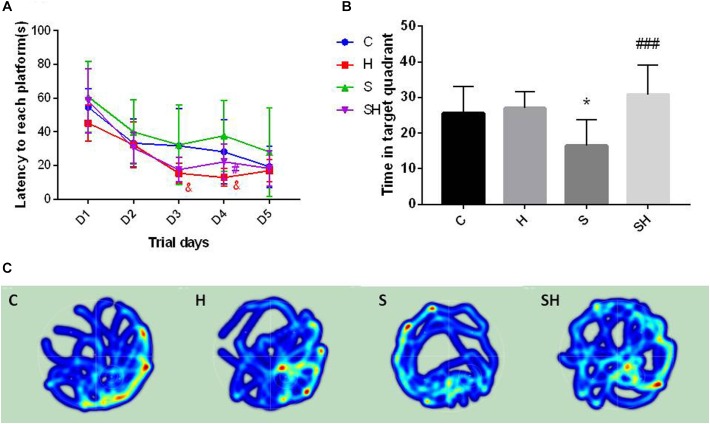
Methods: Postnatal day (P) seven rats were assigned randomly to four groups; (1) group C: non-anesthesia, (2) group H: intraperitoneal hemin (50 mg kg-1) treatment on days 5 and 6, (3) group S: 3% sevoflurane exposure for 4 h, and (4) group SH: hemin treatment + sevoflurane exposure. The expression of neuroglobin in neonatal hippocampus was determined by western blot and immunohistochemistry. Neuroglobin was localized by immunofluorescence. Western blot for the expression of cleaved caspase-3 and TUNEL were used to detect neonatal hippocampal apoptosis, and cytochrome c was used to evaluate mitochondrial function. Drp-1 and Mfn-2 immunoblotting were used to assess mitochondrial dynamics. The Morris water maze test was performed to detect cognitive function in the rats on P30.

Results: Exposure to sevoflurane increased the expression of cleaved caspase-3, cytochrome c, and Drp1 in the neonatal hippocampus and resulted in cognitive deficiency but decreased expression of Mfn2. Hemin reduced apoptosis, improved mitochondrial dynamics and ameliorated the cognitive impairment caused by sevoflurane exposure.

Conclusion: Hemin reduced neuronal apoptosis, improved mitochondrial dynamics and protected against cognitive deficits induced by sevoflurane in neonatal rats. This neuroprotective effect may be achieved by increasing the expression of neuroglobin.

Keywords: hemin; mitochondria; neuroglobin; neurotoxicity; sevoflurane.


Figures

FIGURE 1
FIGURE 1
Hemin inhibits the increase of apoptosis caused by sevoflurane Expression and representative image of cleaved caspase-3 (A) and cytochrome c (B) in the neonatal hippocampus 18 h after sevoflurane exposure. (P< 0.05 vs. group C, ∗∗∗P< 0.001 vs. group C, #P< 0.05 vs. group S, n = 6/group).
FIGURE 2
FIGURE 2
Quantification and detection of TUNEL-positive cells in the hippocampal CA1 region of the neonatal rat brain 18 h after anesthesia Coronal slices for representative TUNEL staining in control (A), H (B), S (C), and SH groups (D). (×400 magnification) (P< 0.05 vs. group C, #P< 0.05 vs. group S, n = 6/group). Scale bar = 50 μm.
FIGURE 3
FIGURE 3
Hemin mitigates the mitochondrial dynamic dysfunction induced by sevoflurane Expression and representative image of Drp1 (A) and Mfn2 (B) in the neonatal hippocampus 18 h after sevoflurane exposure. (P< 0.05 vs. group C, #P< 0.05, ##P< 0.01 vs. group S, n = 6/group).
FIGURE 4
FIGURE 4
Hemin increases the expression of Ngb. (A) Immunohistochemistry of neuroglobin in the neonatal hippocampal CA1 region in each group. Scale bar = 50 μm. (B) Integrated optical density of neuroglobin in the neonatal rat hippocampus. (P< 0.05 vs. group C, ∗∗P< 0.01 vs. group C, n = 6/group). (C) Expression and representative image of neuroglobin in the neonatal hippocampus 18 h after sevoflurane exposure. (P< 0.05 vs. group C, ∗∗∗P< 0.001 vs. group C, n = 6/group).
FIGURE 5
FIGURE 5
Location of neuroglobin Detection of neuroglobin (Ngb) by immunofluorescence double staining in the hippocampus of the neonatal rat brain 18 h after anesthesia in group H. GFAP (red) co-localized with Ngb (green) shown in (A) and NeuN (red) co-localized with Ngb (green) shown in (B). Scale bar = 50 μm.
FIGURE 6
FIGURE 6
Hemin reduces sevoflurane-induced cognitive deficiency The Morris water maze test was used to assess spatial learning and memory ability of 30 day old rats. (A) Escape latency between groups. (&P< 0.05 group H vs. group C, #P< 0.05 vs. group S, n = 9/group) (B) Time of target quadrant (P< 0.05 vs. group C, ###P< 0.001 vs. group S, n = 9/group). (C) Heatmap of time in the target quadrant.

Similar articles

Cited by

References

    1. Amrock L. G., Starner M. L., Murphy K. L., Baxter M. G. (2015). Long-term effects of single or multiple neonatal sevoflurane exposures on rat hippocampal ultrastructure. Anesthesiology 122 87–95. 10.1097/aln.0000000000000477 - DOI - PubMed
    1. Antao S. T., Duong T. T., Aran R., Witting P. K. (2010). Neuroglobin overexpression in cultured human neuronal cells protects against hydrogen peroxide insult via activating phosphoinositide-3 kinase and opening the mitochondrial K(ATP) channel. Antioxid. Redox Signal. 13 769–781. 10.1089/ars.2009.2977 - DOI - PubMed
    1. Baez E., Echeverria V., Cabezas R., Avila-Rodriguez M., Garcia-Segura L. M., Barreto G. E. (2016). Protection by neuroglobin expression in brain pathologies. Front. Neurol. 7:146 10.3389/fneur.2016.00146 - DOI - PMC - PubMed
    1. Boscolo A., Milanovic D., Starr J. A., Sanchez V., Oklopcic A., Moy L., et al. (2013). Early exposure to general anesthesia disturbs mitochondrial fission and fusion in the developing rat brain. Anesthesiology 118 1086–1097. 10.1097/ALN.0b013e318289bc9b - DOI - PMC - PubMed
    1. Burmester T., Weich B., Reinhardt S., Hankeln T. (2000). A vertebrate globin expressed in the brain. Nature 407 520–523. 10.1038/35035093 - DOI - PubMed