. 2019 May 28:12:134.
doi: 10.3389/fnmol.2019.00134. eCollection 2019.

Resilience and Vulnerability to Trauma: Early Life Interventions Modulate Aversive Memory Reconsolidation in the Dorsal Hippocampus

Affiliations

Resilience and Vulnerability to Trauma: Early Life Interventions Modulate Aversive Memory Reconsolidation in the Dorsal Hippocampus

Natividade de Sá Couto-Pereira et al. Front Mol Neurosci. .

Abstract

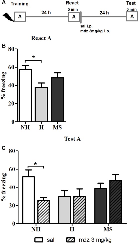
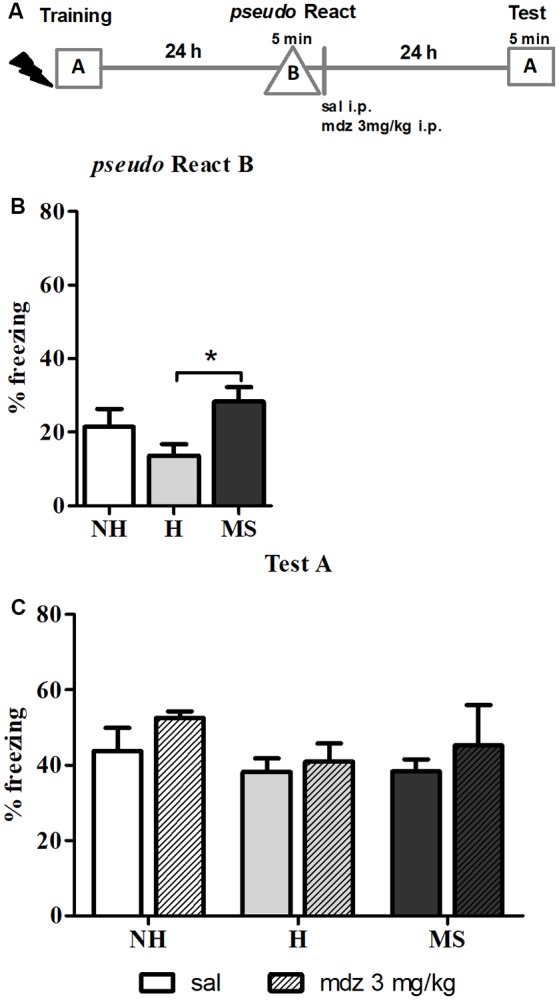
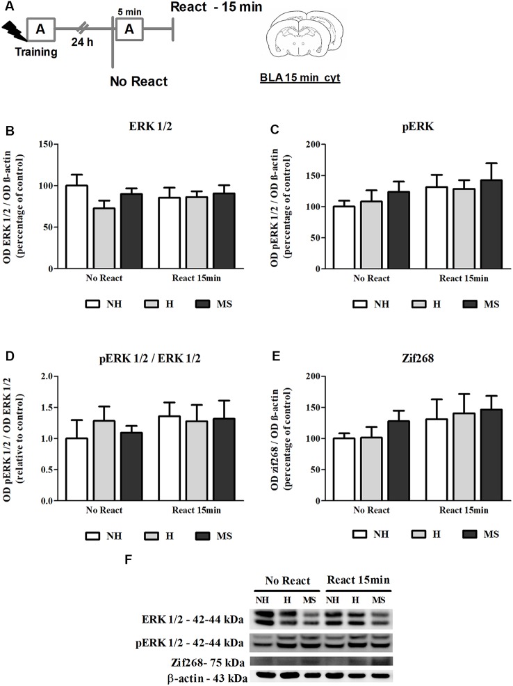
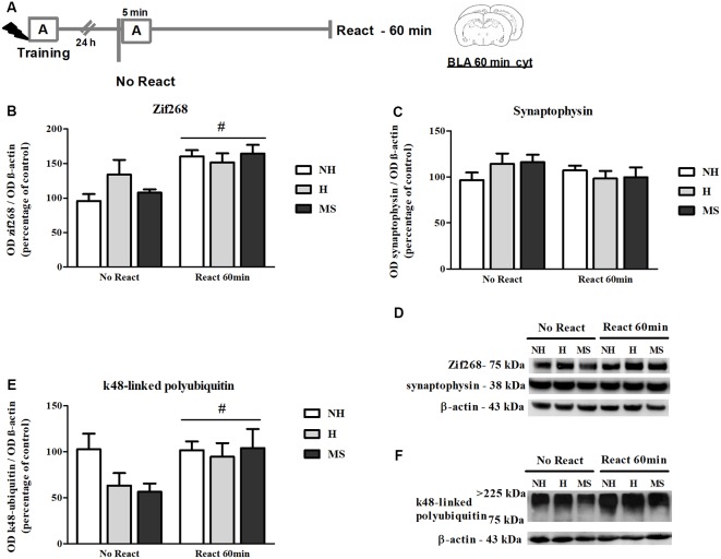
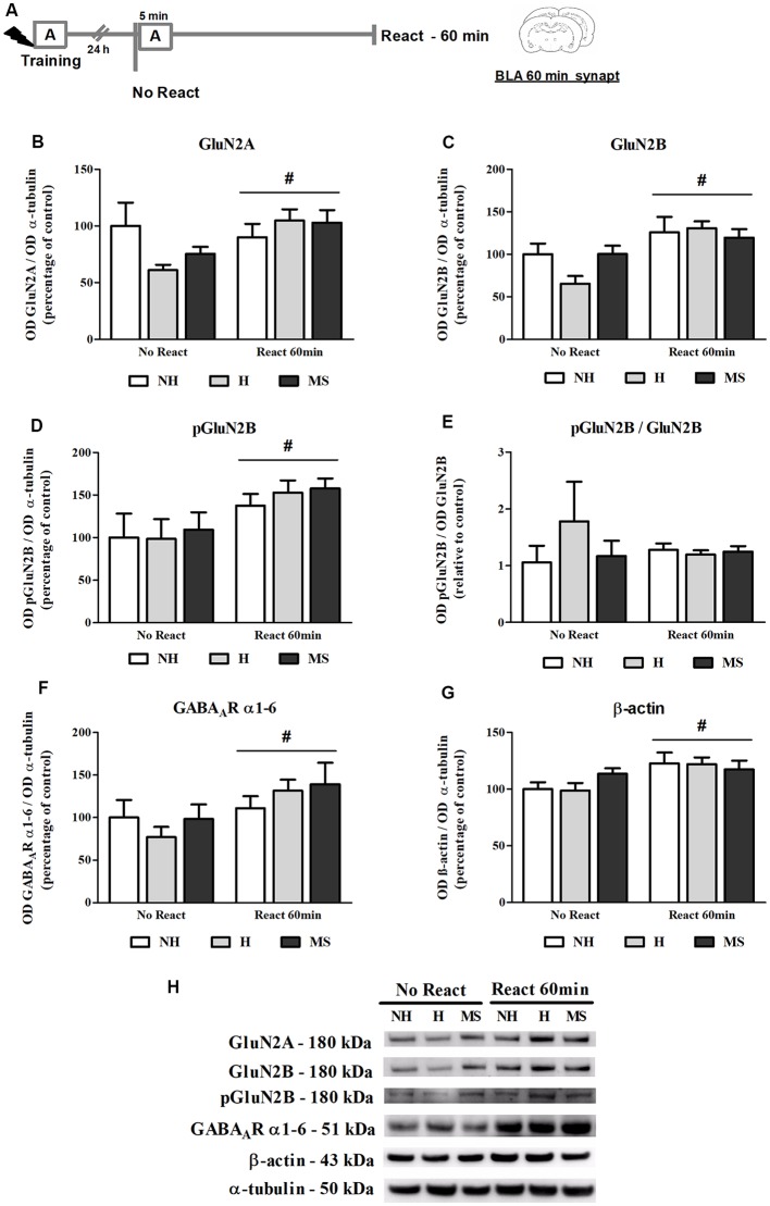
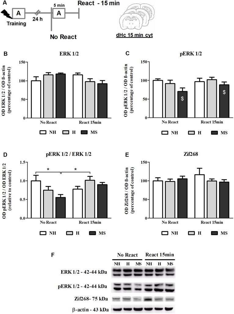
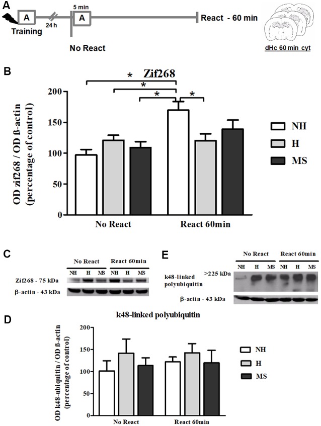
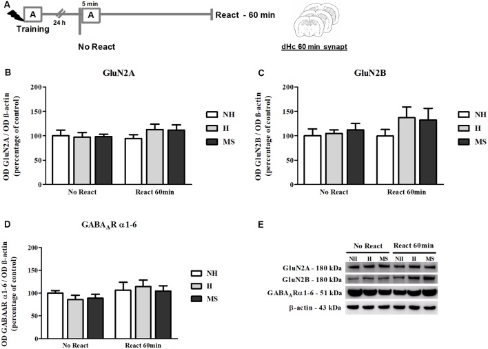
Early life experiences program lifelong responses to stress. In agreement, resilience and vulnerability to psychopathologies, such as posttraumatic stress disorder (PTSD), have been suggested to depend on the early background. New therapies have targeted memory reconsolidation as a strategy to modify the emotional valence of traumatic memories. Here, we used animal models to study the molecular mechanism through which early experiences may later affect aversive memory reconsolidation. Handling (H)-separation of pups from dams for 10 min-or maternal separation (MS) - 3-h separation-were performed from PDN1-10, using non-handled (NH) litters as controls. Adult males were trained in a contextual fear conditioning (CFC) task; 24 h later, a short reactivation session was conducted in the conditioned or in a novel context, followed by administration of midazolam 3 mg/kg i.p. (mdz), known to disturb reconsolidation, or vehicle; a test session was performed 24 h after. The immunocontent of relevant proteins was studied 15 and 60 min after memory reactivation in the dorsal hippocampus (dHc) and basolateral amygdala complex (BLA). Mdz-treated controls (NH) showed decreased freezing to the conditioned context, consistent with reconsolidation impairment, but H and MS were resistant to labilization. Additionally, MS males showed increased freezing to the novel context, suggesting fear generalization; H rats showed lower freezing than the other groups, in accordance with previous suggestions of reduced emotionality facing adversities. Increased levels of Zif268, GluN2B, β-actin and polyubiquitination found in the BLA of all groups suggest that memory reconsolidation was triggered. In the dHc, only NH showed increased Zif268 levels after memory retrieval; also, a delay in ERK1/2 activation was found in H and MS animals. We showed here that reconsolidation of a contextual fear memory is insensitive to interference by a GABAergic drug in adult male rats exposed to different neonatal experiences; surprisingly, we found no differences in the reconsolidation process in the BLA, but the dHc appears to suffer temporal desynchronization in the engagement of reconsolidation. Our results support a hippocampal-dependent mechanism for reconsolidation resistance in models of early experiences, which aligns with current hypotheses for the etiology of PTSD.

Keywords: basolateral amygdala; dorsal hippocampus; fear memory; maternal separation; neonatal handling; reconsolidation.


Figures

Figure 1
Figure 1
Effect of midazolam (mdz) injection after memory reactivation by re-exposure to context A, in adult male rats that were non-handled (NH) or subjected to handling (H) or maternal separation (MS) in the neonatal period. Only NH animals were sensitive to the disrupting effect of mdz on reconsolidation. (A) Schematic diagram of the experimental design. (B) Freezing in context A, in the Reactivation (React) session, n = 17–20/group. (C) Freezing in context A, in the Test session, of animals that received either sal or mdz 3 mg/kg i.p. after the React session, n = 8–10/group. Data are expressed as mean ± standard error of the mean (SEM), as percentage of total session duration. Two-way analysis ofvariance (2w-ANOVA) was used for statistical analyses; *p < 0.05. Statistics results are presented in detail in subsections “Mdz Disrupts Memory Reconsolidation in NH but Not in H and MS Adult Male Rats” and “H Animals Exhibit Less Freezing When Re-exposed to the Conditioned Context.”
Figure 2
Figure 2
The effect of midazolam (mdz) injection after a pseudo-reactivation in context B was tested by re-exposure to context A, in adult male rats that were non-handled (NH) or subjected to handling (H) or maternal separation (MS) in the neonatal period. Mdz effect on reconsolidation is specific to reactivated memories. (A) Schematic diagram of the experimental design. (B) Freezing in context B, in the pseudo Reactivation (React) session, n = 15–17/group. (C) Freezing in context A, in the Test session, of animals that received either sal or mdz 3 mg/kg after the pseudo React B session, n = 7–9/group. Data are expressed as mean ± SEM, in percentage of total session duration. 2w-ANOVA was used for statistical analyses; *p < 0.05. Statistics results are presented in detail in subsections “MS Rats Generalize the Fear Response to Novel Environments” and “Mdz Disrupting Effect on Reconsolidation Requires Properly Reactivated Memories.”
Figure 3
Figure 3
ERK 1/2, pERK 1/2 and Zif268 immunocontent in the basolateral amygdala complex (BLA) cytosolic fraction (cyt) of adult male rats that were non-handled (NH) or subjected to handling (H) or maternal separation (MS) in the neonatal period, 15 min after Reactivation (React) compared to trained animals that were not re-exposed to the training context (No React). No significant differences in ERK 1/2 activation or Zif268 expression were found in the BLA at this time point. (A) Schematic diagram of the experimental design; (B) ERK 1/2; (C) pERK 1/2; (D) calculated ratio of pERK 1/2 per ERK 1/2 immunocontent; (E) Zif268; (F) representative Western blot bands. Data are expressed as mean ± SEM. n = 5–7/group. 2w-ANOVA was used for statistical analyses. Statistics results are presented in detail in subsection “ERK 1/2 Activity and Zif268 Levels Were not Changed in the BLA 15 min After Aversive Memory Reactivation.”
Figure 4
Figure 4
Zif268, synaptophysin and k48-linked polyubiquitinated proteins immunocontent in the basolateral amygdala complex (BLA) cytosolic fraction (cyt) of adult male rats that were non-handled (NH) or subjected to handling (H) or maternal separation (MS) in the neonatal period, 60 min after Reactivation (React) compared to trained animals that were not re-exposed to the training context (No React). Memory reactivation induced a significant increase in Zif268 and polyubiquitinated proteins in the BLA cyt of all groups. (A) Schematic diagram of the experimental design; (B) Zif268; (C) synaptophysin; (D) representative Western blot bands; (E) k48-linked polyubiquitin proteins; (F) representative Western blot bands. Data are expressed as mean ± SEM. n = 5–8/group. 2w-ANOVA was used for statistical analyses; #p < 0.05 (main effect of reactivation). Statistics results are presented in detail in subsections “Zif268 Levels Increase in the BLA, 60 min After Aversive Memory Reactivation,” “Memory Reactivation Induces Changes in Receptor Composition at the BLA Synapses” and “Synaptic NMDA and GABAAR Subunits Were not Changed by Memory Reactivation in the dHc.”
Figure 5
Figure 5
NMDA receptor (NMDAR) subunits GluN2A and GluN2B (total and phosphorylated), GABAAR α1–6 subunits and β-actin immunocontent in the basolateral amygdala complex (BLA) synaptosome membrane fraction (synapt) of adult male rats that were non-handled (NH) or subjected to handling (H) or maternal separation (MS) in the neonatal period, 60 min after Reactivation (React) compared to trained animals that were not re-exposed to the training context (No React). Memory reactivation induced a significant increase in all receptor subunits and also β-actin in the BLA synapt of all groups. (A) Schematic diagram of the experimental design; (B) GluN2A; (C) GluN2B; (D) pGluN2B; (E) calculated ratio of pGluN2B per GluN2B immunocontent; (F) GABAAR α1–6 subunits; (G) β-actin; (H) representative Western blot bands. Data are expressed as mean ± SEM. n = 5–7/group. 2w-ANOVA was used for statistical analyses. #p < 0.05 (main effect of reactivation). Statistics results are presented in detail in subsection “Memory Reactivation Induces Changes in Receptor Composition at the BLA Synapses.”
Figure 6
Figure 6
NMDAR subunits GluN2A and GluN2B and GABAAR α1–6 subunits immunocontent in the basolateral amygdala complex (BLA) cytosolic fraction (cyt) of adult male rats that were non-handled (NH) or subjected to handling (H) or maternal separation (MS) in the neonatal period. (A) Schematic diagram of the experimental design; (B) GluN2A; (C) GluN2B; (D) GABAAR α1–6 subunits; (E) representative Western blot bands. Data are expressed as mean ± SEM. n = 6–8/group. 1w-ANOVA was used for statistical analyses. *p < 0.05. Statistics results are presented in detail in subsection “Memory Reactivation Induces Changes in Receptor Composition at the BLA Synapses.”
Figure 7
Figure 7
ERK 1/2, pERK 1/2 and Zif268 immunocontent in the dorsal hippocampus (dHc) cytosolic fraction (cyt) of adult male rats that were non-handled (NH) or subjected to handling (H) or maternal separation (MS) in the neonatal period, 15 min after Reactivation (React) compared to trained animals that were not re-exposed to the training context (No React). Activation of ERK 1/2 in the dHc appears to be delayed, particularly in MS rats. (A) Schematic diagram of the experimental design; (B) ERK 1/2; (C) pERK 1/2; (D) calculated ratio of pERK 1/2 per ERK 1/2 immunocontent; (E) Zif268; (F) representative Western blot bands. Data are expressed as mean ± SEM. n = 5–8/group. 2w-ANOVA was used for statistical analyses. *p < 0.05; $p < 0.05 (main effect of neonatal intervention). Statistics results are presented in detail in subsection “Neonatal Interventions Change ERK 1/2 Activation in the dHc, 15 min After Aversive Memory Reactivation.”
Figure 8
Figure 8
Zif268 and k48-linked polyubiquitinated proteins immunocontent in the dHc cytosolic fraction (cyt) of adult male rats that were non-handled (NH) or subjected to handling (H) or maternal separation (MS) in the neonatal period, 60 min after Reactivation (React) compared to trained animals that were not re-exposed to the training context (No React). Only NH rats exhibited the expected increase in Zif268 in the dHc induced by memory reactivation. (A) Schematic diagram of the experimental design; (B) Zif268; (C) representative Western blot bands; (D) k48-linked polyubiquitin proteins; (E) representative Western blot bands. Data are expressed as mean ± SEM. n = 6–8/group. 2w-ANOVA was used for statistical analyses; *p < 0.05. Statistics results are presented in detail in subsections “Zif268 Levels Increase in the dHc of NH, but Not H or MS, 60 min After Aversive Memory Reactivation” and “k48-Linked Polyubiquitin Levels Were Increased by Reactivation in the BLA, but Not in dHc.”
Figure 9
Figure 9
NMDAR subunits GluN2A and GluN2B and GABAAR α1–6 subunits immunocontent in the dHc synaptosome membrane fraction (synapt) of adult male rats that were non-handled (NH) or subjected to handling (H) or maternal separation (MS) in the neonatal period, 60 min after Reactivation (React) compared to trained animals that were not re-exposed to the training context (No React). No significant changes in N-Methyl-D-aspartate (NMDA) or GABAAR subunit composition were found in the dHc. (A) Schematic diagram of the experimental design; (B) GluN2A; (C) GluN2B; (D) GABAAR α1–6 subunits; (E) representative Western blot bands. Data are expressed as mean ± SEM. n = 5–7/group. 2w-ANOVA was used for statistical analyses. Statistics results are presented in detail in subsection “Synaptic NMDA and GABAAR Subunits Were not Changed by Memory Reactivation in the dHc.”

Similar articles

Cited by

References

    1. Abdallah C. G., Wrocklage K. M., Averill C. L., Akiki T., Schweinsburg B., Roy A., et al. . (2017). Anterior hippocampal dysconnectivity in posttraumatic stress disorder: a dimensional and multimodal approach. Transl. Psychiatry 7:e1045. 10.1038/tp.2017.12 - DOI - PMC - PubMed
    1. Agustina López M., Jimena Santos M., Cortasa S., Fernández R. S., Carbó Tano M., Pedreira M. E. (2016). Different dimensions of the prediction error as a decisive factor for the triggering of the reconsolidation process. Neurobiol. Learn. Mem. 136, 210–219. 10.1016/j.nlm.2016.10.016 - DOI - PubMed
    1. Akirav I., Maroun M. (2013). Stress modulation of reconsolidation. Psychopharmacology 226, 747–761. 10.1007/s00213-012-2887-6 - DOI - PubMed
    1. Andersen S. L., Teicher M. H. (2004). Delayed effects of early stress on hippocampal development. Neuropsychopharmacology 29, 1988–1993. 10.1038/sj.npp.1300528 - DOI - PubMed
    1. Arnett M. G., Pan M. S., Doak W., Cyr P. E. P., Muglia L. M., Muglia L. J. (2015). The role of glucocorticoid receptor-dependent activity in the amygdala central nucleus and reversibility of early-life stress programmed behavior. Transl. Psychiatry 5:e542. 10.1038/tp.2015.35 - DOI - PMC - PubMed