

Quantifying How Staining Methods Bias Measurements of Neuron Morphologies
- PMID: 31191283
- PMCID: PMC6541099
- DOI: 10.3389/fninf.2019.00036
Quantifying How Staining Methods Bias Measurements of Neuron Morphologies
Abstract
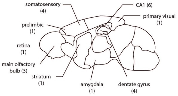
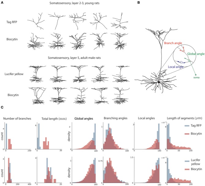
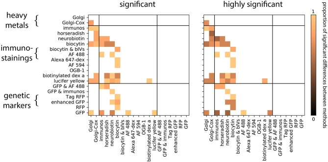
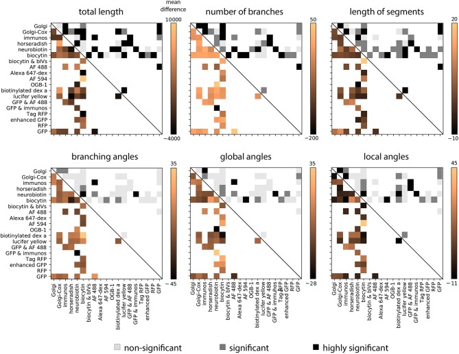
The process through which neurons are labeled is a key methodological choice in measuring neuron morphology. However, little is known about how this choice may bias measurements. To quantify this bias we compare the extracted morphology of neurons collected from the same rodent species, experimental condition, gender distribution, age distribution, brain region and putative cell type, but obtained with 19 distinct staining methods. We found strong biases on measured features of morphology. These were largest in features related to the coverage of the dendritic tree (e.g., the total dendritic tree length). Understanding measurement biases is crucial for interpreting morphological data.
Keywords: dendritic morphology; fluorescence microscopy; golgi method; immunostaining; neuroinformatics; rodent neuroanatomy; staining method.
Figures
Similar articles
-
Morphological study of the rostral interstitial nucleus of the medial longitudinal fasciculus in the monkey, Macaca mulatta, by Nissl, Golgi, and computer reconstruction and rotation methods.J Comp Neurol. 1994 Sep 1;347(1):47-63. doi: 10.1002/cne.903470105. J Comp Neurol. 1994. PMID: 7528228
-
A Confocal Reflection Super-Resolution Technique to Image Golgi-Cox Stained Neurons.J Microsc. 2019 Aug;275(2):115-130. doi: 10.1111/jmi.12821. Epub 2019 Jul 11. J Microsc. 2019. PMID: 31237354 Free PMC article.
-
Generation, description and storage of dendritic morphology data.Philos Trans R Soc Lond B Biol Sci. 2001 Aug 29;356(1412):1131-45. doi: 10.1098/rstb.2001.0905. Philos Trans R Soc Lond B Biol Sci. 2001. PMID: 11545695 Free PMC article. Review.
-
A Quantitative Golgi Study of Dendritic Morphology in the Mice Striatal Medium Spiny Neurons.Front Neuroanat. 2017 Apr 28;11:37. doi: 10.3389/fnana.2017.00037. eCollection 2017. Front Neuroanat. 2017. PMID: 28503136 Free PMC article.
-
The need for integrating neuronal morphology databases and computational environments in exploring neuronal structure and function.Anat Embryol (Berl). 2001 Oct;204(4):255-65. doi: 10.1007/s004290100197. Anat Embryol (Berl). 2001. PMID: 11720232 Review.
Cited by
-
A Systematic Evaluation of Interneuron Morphology Representations for Cell Type Discrimination.Neuroinformatics. 2020 Oct;18(4):591-609. doi: 10.1007/s12021-020-09461-z. Neuroinformatics. 2020. PMID: 32367332 Free PMC article.
-
Exploring the relationship between epigenetic DNA methylation and cardiac fibrosis through Raman microspectroscopy.Am J Physiol Cell Physiol. 2023 Jul 1;325(1):C332-C343. doi: 10.1152/ajpcell.00209.2023. Epub 2023 Jun 19. Am J Physiol Cell Physiol. 2023. PMID: 37335025 Free PMC article.
-
Prevalence and practices of immunofluorescent cell image processing: a systematic review.Front Cell Neurosci. 2023 Jul 20;17:1188858. doi: 10.3389/fncel.2023.1188858. eCollection 2023. Front Cell Neurosci. 2023. PMID: 37545881 Free PMC article.
-
Comprehensive Estimates of Potential Synaptic Connections in Local Circuits of the Rodent Hippocampal Formation by Axonal-Dendritic Overlap.J Neurosci. 2021 Feb 24;41(8):1665-1683. doi: 10.1523/JNEUROSCI.1193-20.2020. Epub 2020 Dec 23. J Neurosci. 2021. PMID: 33361464 Free PMC article.
-
Accelerating the continuous community sharing of digital neuromorphology data.FASEB Bioadv. 2024 Jun 17;6(7):207-221. doi: 10.1096/fba.2024-00048. eCollection 2024 Jul. FASEB Bioadv. 2024. PMID: 38974113 Free PMC article.
References
LinkOut - more resources
Full Text Sources