. 2019 May 29:10:1177.
doi: 10.3389/fmicb.2019.01177. eCollection 2019.

Differential Expression of Three Cryptosporidium Species-Specific MEDLE Proteins

Affiliations

Differential Expression of Three Cryptosporidium Species-Specific MEDLE Proteins

Jiayuan Su et al. Front Microbiol. .

Abstract

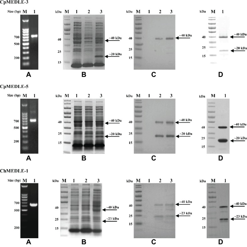
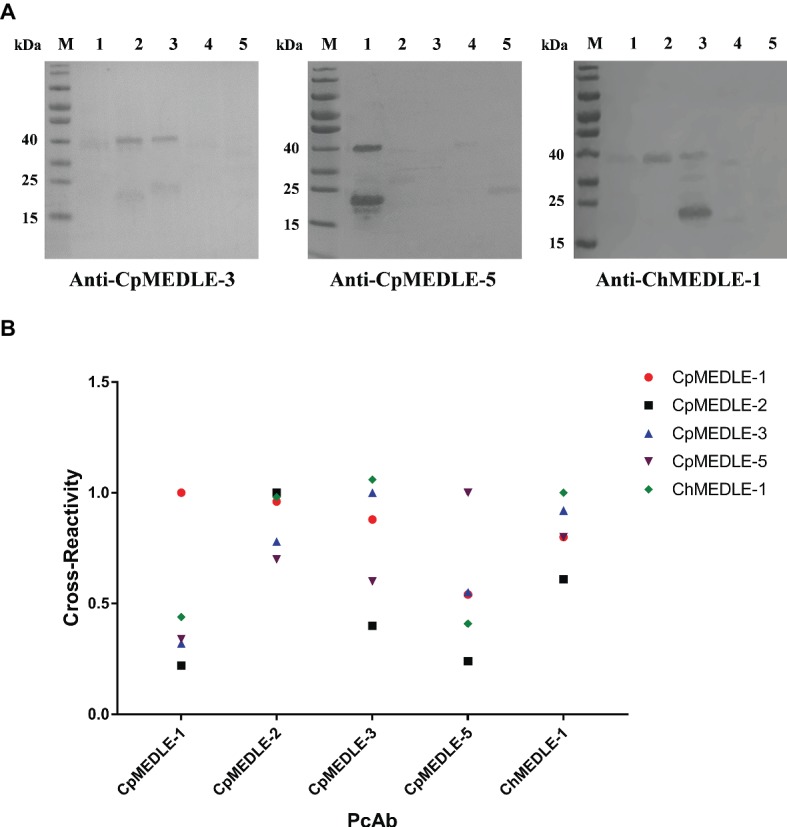
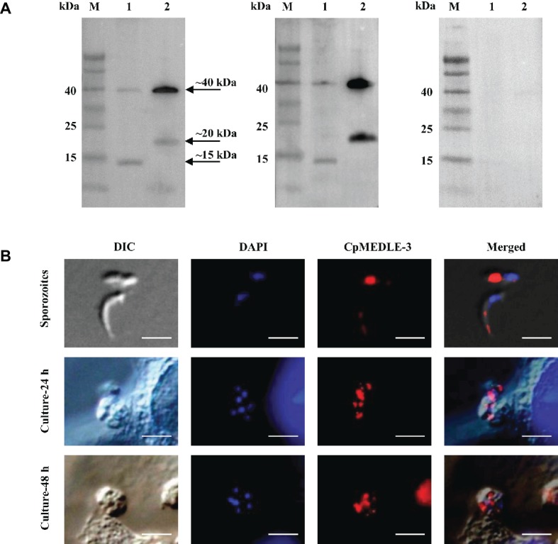
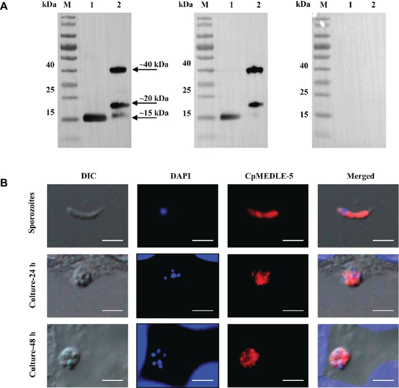
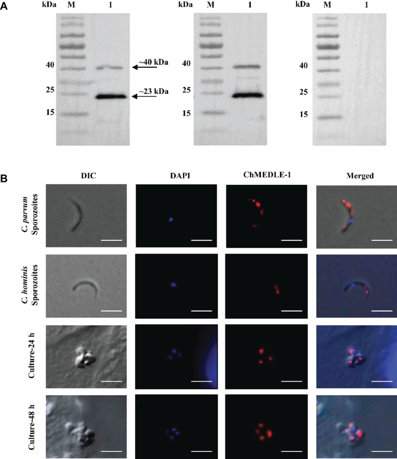
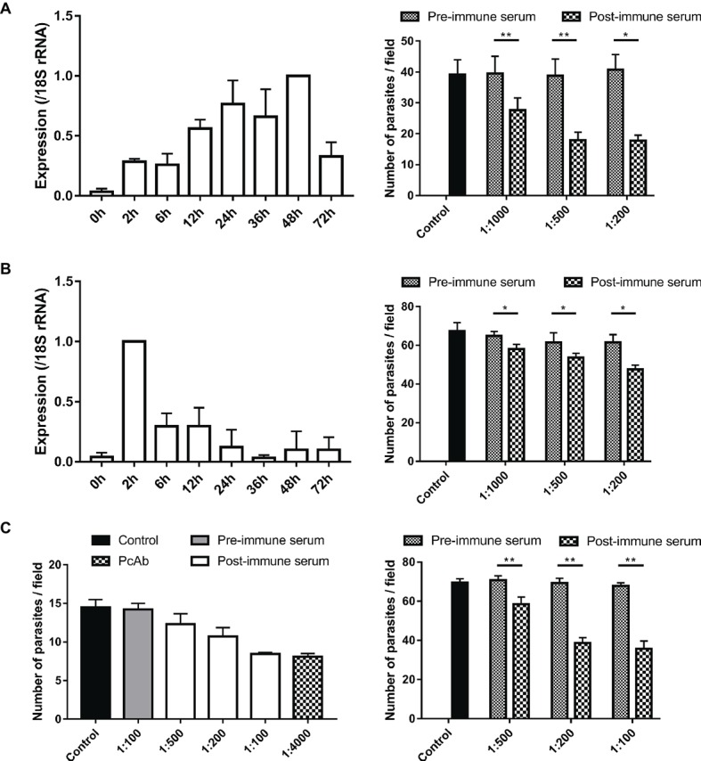
Cryptosporidium parvum and Cryptosporidium hominis share highly similar proteomes, with merely ~3% divergence in overall nucleotide sequences. Cryptosporidium-specific MEDLE family is one of the major differences in gene content between the two species. Comparative genomic analysis indicated that MEDLE family may contribute to differences in host range among Cryptosporidium spp. Previous studies have suggested that CpMEDLE-1 encoded by cgd5_4580 and CpMEDLE-2 encoded by cgd5_4590 are potentially involved in the invasion of C. parvum. In this study, we expressed in Escherichia coli, the C. hominis-specific member of the MEDLE protein family, ChMEDLE-1 encoded by chro.50507, and two C. parvum-specific members, CpMEDLE-3 encoded by cgd5_4600 and CpMEDLE-5 encoded by cgd6_5480. Quantitative PCR, immunofluorescence staining and in vitro neutralization assay were conducted to assess their biologic characteristics. The expression of the cgd5_4600 gene was high during 12-48 h of the in vitro culture, while the expression of cgd6_5480 was the highest at 2 h. ChMEDLE-1 and CpMEDLE-3 proteins were mostly located in the anterior and mid-anterior region of sporozoites and merozoites, whereas CpMEDLE-5 was expressed over the entire surface of these invasive stages. Polyclonal antibodies against MEDLE proteins had different neutralization efficiency, reaching approximately 50% for ChMEDLE-1 and 60% for CpMEDLE-3, but only 20% for CpMEDLE-5. The differences in protein and gene expression and neutralizing capacity indicated the MEDLE proteins may have different roles during Cryptosporidium invasion and growth.

Keywords: Cryptosporidium hominis; Cryptosporidium parvum; MEDLE family; growth; invasion.


Figures

Figure 1
Figure 1
Production and purification of recombinant MEDLE proteins CpMEDLE-3 and CpMEDLE-5 from Cryptosporidium parvum and ChMEDLE-1 from Cryptosporidium hominis. (A) PCR amplification of the target gene in genomic DNA. Lane M: molecular markers; lane 1: PCR product. (B) Expression of recombinant MEDLE protein in E. coli BL21 (DE3) as revealed by SDS-PAGE analysis. Lane M: molecular weight markers; lane 1: lysate from bacteria culture transformed with the recombinant plasmid without IPTG induction; lane 2: lysate from similar bacteria culture induced by IPTG for 2 h; lane 3: lysate from bacteria culture induced by IPTG for 8 h, with the expected product indicated by an arrow. (C) Western blot analysis of the recombinant protein. Lane M: molecular weight markers; lane 1: lysate from bacteria culture transformed with recombinant plasmid without IPTG induction; lane 2: supernatant from IPTG-induced bacterial culture; lane 3: cell lysate from IPTG-induced bacterial culture. (D) Purification of recombinant proteins. Lane M: molecular weight markers; lane 1: purified recombinant proteins from Ni-NTA affinity chromatography.
Figure 2
Figure 2
Cross-reactivity of polyclonal antibodies (PcAb) among MEDLE proteins. (A) Western blot analysis of cross-reactivity among MEDLE proteins, using 1 μg of CpMEDLE-5 (lane 1), CpMEDLE-3 (lane 2), ChMEDLE-1 (lane 3), CpMEDLE-1 (lane 4) and CpMEDLE-2 (lane 5) and PcAb to CpMEDLE-3 (left panel), CpMEDLE-5 (middle panel) and ChMEDLE-1 (right panel). (B) ELISA analysis of polyclonal antibodies to each MEDLE protein, with data being normalized using the relative absorbance of each antibody to the corresponding protein.
Figure 3
Figure 3
Expression of CpMEDLE-3 in sporozoites and developmental stages of Cryptosporidium parvum. (A) Western blots analysis of native protein from C. parvum sporozoites for CpMEDLE-3, using polyclonal antibodies (left panel), post-immune sera (middle panel) and pre-immune sera (right panel). Lane M: molecular weight markers; lane 1: crude protein extracted from sporozoites; lane 2: purified CpMEDLE-3 protein. (B) Expression of CpMEDLE-3 on C. parvum sporozoites (top panel) and intracellular developmental stages in HCT-8 cell cultures at 24 h (middle panel) and 48 h (bottom panel). The images were taken under differential interference contrast (DIC), with nucleus counter-stained with 4′,6-diamidino-2-phenylindole (DAPI), parasites stained by immunofluorescence with Alexa 594-labled CpMEDLE-3 (CpMEDLE-3), and superimposition of the three images (Merged). Bars = 5 μm.
Figure 4
Figure 4
Expression of CpMEDLE-5 in sporozoites and developmental stages of Cryptosporidium parvum. (A) Western blots analysis of native protein from C. parvum sporozoites for CpMEDLE-5, using polyclonal antibodies (left panel), post-immune sera (middle panel) and pre-immune sera (right panel). Lane M: molecular weight markers; lane 1: crude protein extracted from sporozoites; lane 2: purified CpMEDLE-5 protein. (B) Expression of CpMEDLE-5 on C. parvum sporozoites (top panel) and intracellular developmental stages in HCT-8 cell cultures at 24 h (middle panel) and 48 h (bottom panel). The images were taken under differential interference contrast (DIC), with nucleus counter-stained with 4′,6-diamidino-2-phenylindole (DAPI), parasites stained by immunofluorescence with Alexa 594-labled CpMEDLE-5 (CpMEDLE-5), and superimposition of the three images (Merged). Bars = 5 μm.
Figure 5
Figure 5
Expression of ChMEDLE-1. (A) Western blots analysis of ChMEDLE-1 recombinant protein, using polyclonal antibodies (left panel), post-immune sera (middle panel) and pre-immune sera (right panel). Lane M: molecular weight markers; lane 1: purified ChMEDLE-1 protein. (B) Expression of ChMEDLE-1 on C. parvum sporozoites (top panel), C. hominis sporozoites (the second panel) and intracellular developmental stages of C. hominis in HCT-8 cell cultures at 24 h (the third panel) and 48 h (bottom panel). The images were taken under differential interference contrast (DIC), with nucleus counter-stained with 4′,6-diamidino-2-phenylindole (DAPI), parasites stained by immunofluorescence with Alexa 594-labled ChMEDLE-1 (ChMEDLE-1), and superimposition of the three images (Merged). Bars = 5 μm.
Figure 6
Figure 6
Assessment of biological functions of MEDLE proteins. (A) Expression levels of the cgd5_4600 gene in developmental stages of C. parvum (left panel) and neutralization efficiency of immune sera against CpMEDLE-3 on C. parvum invasion (right panel). The expression level of the cgd5_4600 gene in C. parvum culture was determined by qPCR at various time points, with data being normalized with data from the expression of the Cp18S rRNA. Neutralization efficiency of post-immune sera against CpMEDLE-3 on C. parvum invasion was measured in HCT-8 cell culture. Data presented are mean ± SD from three independent experiments for both the expression and neutralization studies. (B) Expression levels of the cgd6_5480 gene in developmental stages of C. parvum (left panel) and neutralization efficiency of immune sera against CpMEDLE-5 on C. parvum invasion (right panel). The expression level of the cgd6_5480 gene in C. parvum culture was determined by qPCR at various time points, with data being normalized with data from the expression of the Cp18S rRNA. Neutralization efficiency of immune sera against CpMEDLE-5 on C. parvum invasion was measured in HCT-8 cell culture. Data presented are mean ± SD from three independent experiments for both the expression and neutralization studies. (C) Neutralization efficiency of polyclonal antibodies (PcAb) and immune sera against ChMEDLE-1 on C. hominis (left panel) and C. parvum (right panel) invasion. Cultures were treated with PcAb, immune sera, or pre-immune sera. Data presented are mean ± SD from two (for C. hominis) or three (for C. parvum) independent experiments. The statistical significance of differences between treatment groups is indicated for neutralization studies of all three MEDLE proteins above the bars (*p < 0.05; **p < 0.01).

Similar articles

Cited by

References

    1. Abrahamsen M. S., Templeton T. J., Enomoto S., Abrahante J. E., Zhu G., Lancto C. A., et al. . (2004). Complete genome sequence of the apicomplexan, Cryptosporidium parvum. Science 304, 441–445. 10.1126/science.1094786, PMID: - DOI - PubMed
    1. Arrowood M. J., Donaldson K. (1996). Improved purification methods for calf-derived Cryptosporidium parvum oocysts using discontinuous sucrose and cesium chloride gradients. J. Eukaryot. Microbiol. 43:89S. 10.1111/j.1550-7408.1996.tb05015.x, PMID: - DOI - PubMed
    1. Beverley S. M. (2015). Parasitology: CRISPR for Cryptosporidium. Nature 523, 413–414. 10.1038/nature14636, PMID: - DOI - PMC - PubMed
    1. Bhalchandra S., Cardenas D., Ward H. D. (2018). Recent breakthroughs and ongoing limitations in Cryptosporidium research. F1000Res. 7:1380. 10.12688/f1000research.15333.1, PMID: - DOI - PMC - PubMed
    1. Cai X., Woods K. M., Upton S. J., Zhu G. (2005). Application of quantitative real-time reverse transcription-PCR in assessing drug efficacy against the intracellular pathogen Cryptosporidium parvum in vitro. Antimicrob. Agents Chemother. 49, 4437–4442. 10.1128/AAC.49.11.4437-4442.2005, PMID: - DOI - PMC - PubMed