. 2019 May 29:10:1202.
doi: 10.3389/fmicb.2019.01202. eCollection 2019.

Seneca Valley Virus 2C and 3Cpro Induce Apoptosis via Mitochondrion-Mediated Intrinsic Pathway

Affiliations

Seneca Valley Virus 2C and 3Cpro Induce Apoptosis via Mitochondrion-Mediated Intrinsic Pathway

Tingting Liu et al. Front Microbiol. .

Abstract

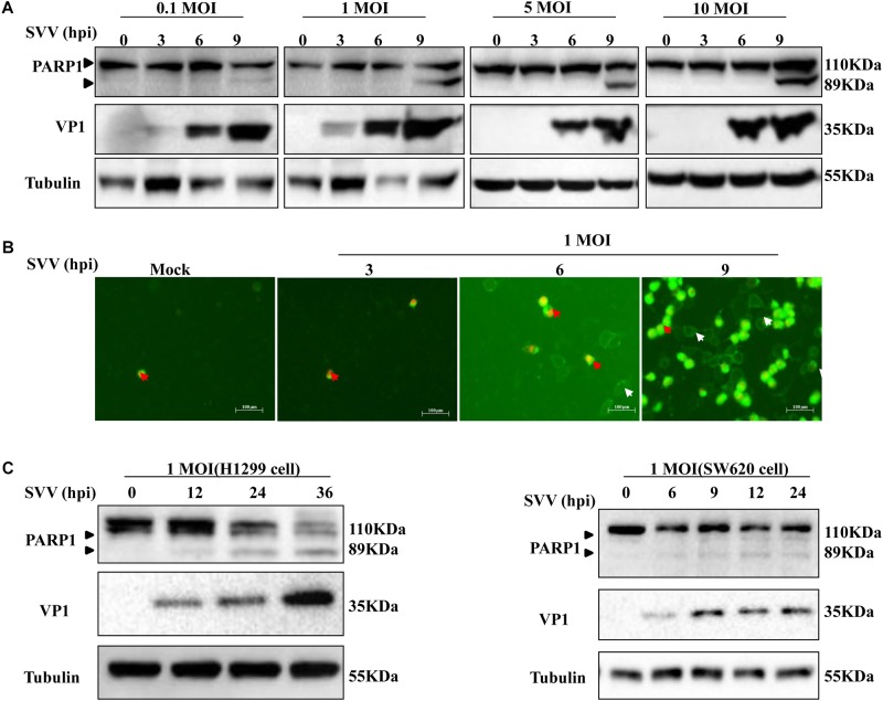
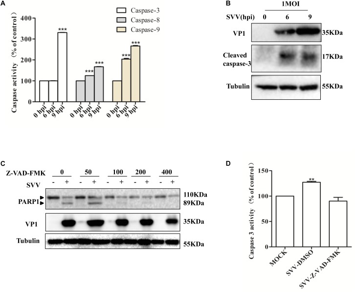
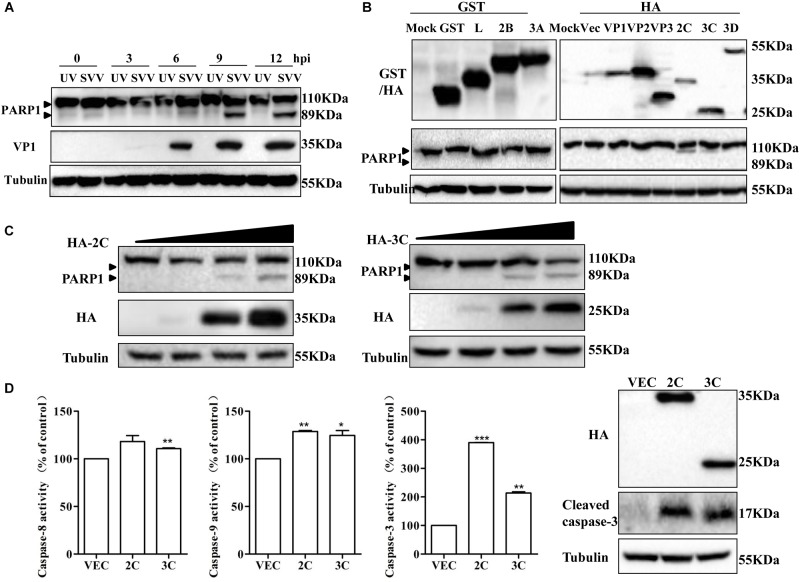
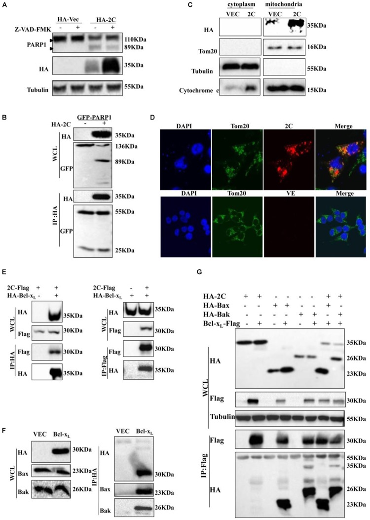
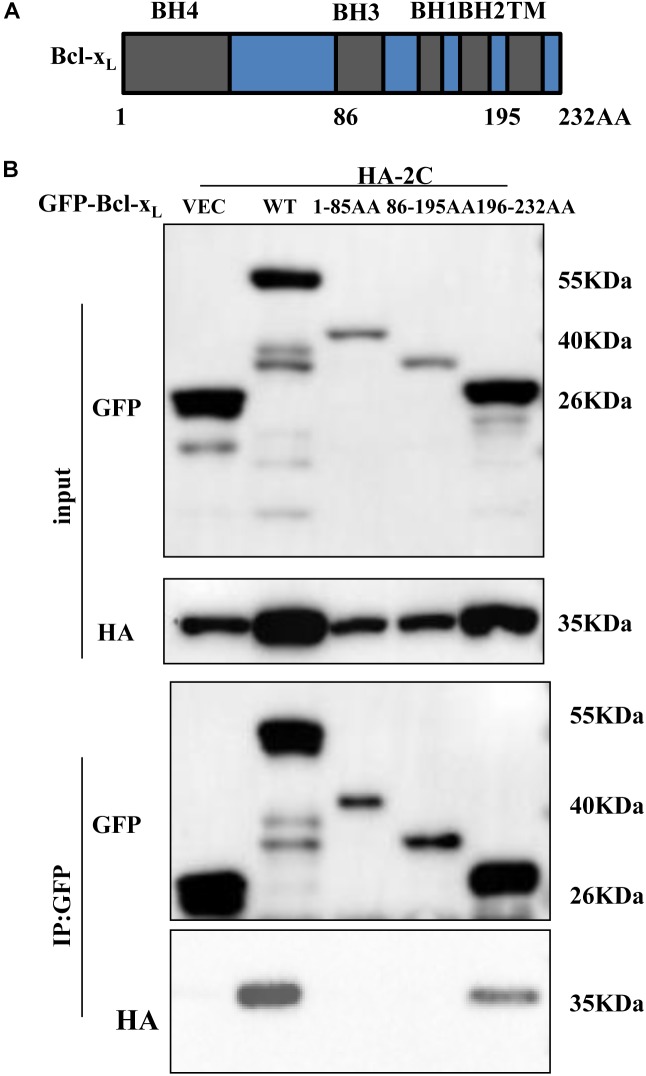
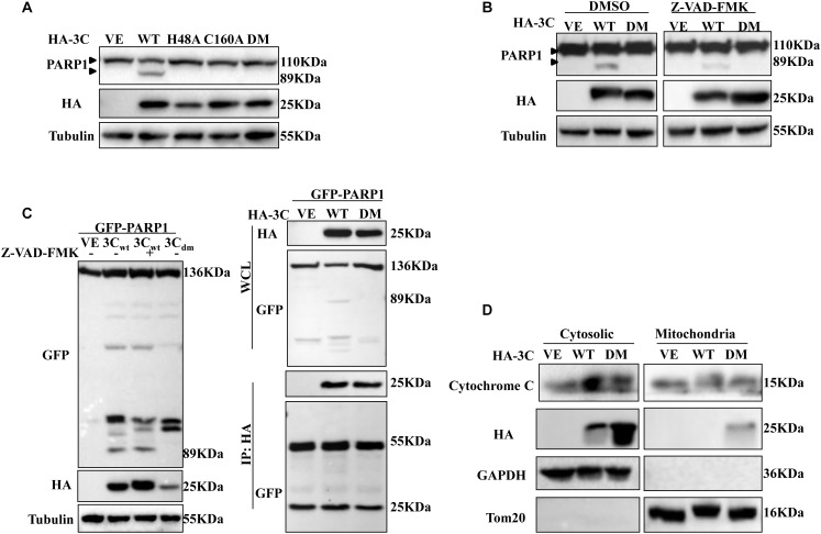
Seneca Valley virus (SVV) is the only member of the genus Senecavirus of the Picornaviridae family. SVV can selectively infect and lyse tumor cells with neuroendocrine features and is used as an oncolytic virus for treating small-cell lung cancers. However, the detailed mechanism underlying SVV-mediated destruction of tumor cells remains unclear. In this study, we found that SVV can increase the proportion of apoptotic 293T cells in a dose- and time-dependent manner. SVV-induced apoptosis was initiated via extrinsic and intrinsic pathways through activation of caspase-3, the activity of which could be attenuated by a pan-caspase inhibitor (Z-VAD-FMK). We confirmed that SVV 2C and 3Cpro play critical roles in SVV-induced apoptosis. The SVV 2C protein was located solely in the mitochondria and activated caspase-3 to induce apoptosis. SVV 3Cpro induced apoptosis through its protease activity, which was accompanied by release of cytochrome C into the cytoplasm, but did not directly cleave PARP1.

Keywords: 2C; 3Cpro; apoptosis; caspase-3; seneca valley virus (SVV).


Figures

FIGURE 1
FIGURE 1
SVV induces cell apoptosis. (A) 293T cells were challenged with SVV at different MOIs (0.1, 1, 5, and 10), then the cells were harvested at indicated times and lysed, analyzed by western blotting with the indicated antibodies. (B) 293T cells were treated with SVV at an MOI for the indicated time, then the cells were stained with Annexin V-FITC/PI and observed with the fluorescence microscope. (C) H1299 cells and SW620 cells were infected with SVV at one MOI for the indicated time, and cleavage of PARP1 was determined by western blotting analysis.
FIGURE 2
FIGURE 2
SVV induces cells apoptosis through both extrinsic and intrinsic pathways. (A) SVV infected 293T cells at an MOI for the indicated time, then the activities of the caspase-8, caspase-9, and caspase-3 were detected by the corresponding Caspase Activity Assay Kit. (B) SVV infected 293T cells at an MOI for the indicated time, then the cell lysates were analyzed by western blotting to detect the expression levels of cleaved caspase-3 bands. (C) Cell lysates from mock-treated or SVV-infected 293T cells in the presence of different concentrations of caspase inhibitor Z-VAD-FMK (0, 50, 100, 200, and 400 μM) were subjected to western blotting analysis with the indicated antibodies. (D) The activity of caspase-3 of the cells infected with SVV at one MOI in the absence or presence of Z-VAD-FMK at 50 μM by the Caspase-3 Activity Assay Kit ( 0.01 < P < 0.05; ∗∗ P < 0.01; ∗∗∗ P < 0.001).
FIGURE 3
FIGURE 3
SVV 2C and 3Cpro induce 293T cells apoptosis via activating caspase-3. (A) SVV were inactivated with UV, then 293T cells were challenged with the inactivated SVV and SVV at one MOI at the indicated time points post-infection, the cells were collected and lysed to be subjected to western blotting with the indicated antibodies. (B) 293T cells were transfected with the indicated plasmids expressing GST-tag or HA-tag viral proteins or the corresponding empty vector plasmids. The expression of these viral proteins and cleavage of PARP1 were detected by western blotting at 24 hpt. (C) 293T cells were transfected with different doses of HA-2C or HA-3C expressing plasmids for 24 h. The expression of corresponding proteins was determined by western blotting. (D) 293T cells were transfected with the indicated plasmids expressing 2C or 3C proteins, then the cells were harvested to measured the activities of the caspase-8, caspase-9 and caspase-3 by the corresponding Caspase Activity Assay Kit. The cell lysates were analyzed by western blotting to detect the expression levels of cleaved caspase-3 bands (0.01 < P < 0.05; ∗∗P < 0.01; ∗∗∗P < 0.001).
FIGURE 4
FIGURE 4
2C protein is located in the mitochondria and interacts with Bcl-xL. (A) 293T cells were transfected with HA-2C in the absence or presence of Z-VAD-FMK (50 μM) for 24 h. The cells lysates were analyzed by western blotting to detect the expression levels of cleaved PARP1 bands. (B) 293T cells were cotransfected with HA-2C and GFP-PARP1 for 24 h and then subjected to immunoprecipitation (IP) with anti-HA antibody. The immunoprecipitates were analyzed by western blotting with the indicated antibodies. (C) Western blotting analysis of 2C protein in the cytosol and mitochondrial fractions of cells expressing 2C protein. Cytosolic and mitochondrial fractions were separated, and equal amounts of proteins from each fraction were immunoblotted with anti-GAPDH, anti-cytochrome C, anti-HA, or anti-Tom20 antibody (internal control of mitochondrial fraction). (D) An experiment indicating that 2C was located in the mitochondria. Cells were transfected with HA-2C (red arrow) or empty vector. At 24 h posttransfection, the cells were fixed and analyzed by confocal fluorescence microscopy. Tom20 served as a mitochondrial marker and was stained with anti-Tom20 (green) antibody, and then imaged by confocal microscopy. (E) 293T cells were cotransfected with HA-Bcl-xL and 2C-Flag for 24 h. Lysates were immunoprecipitated by anti-HA or anti-Flag antibody as indicated. (F) 293T cells were transfected with HA-Bcl-xL for 24 h and then subjected to immunoprecipitation with anti-HA, anti-Bak, or anti-Bax antibody. (G) The indicated plasmids were cotransfected into 293T cells for 24 h and analyzed by Co-IP using the indicated antibody.
FIGURE 5
FIGURE 5
2C protein interacts with the C-terminal region (196-232AA) of Bcl-xL protein. (A) Schematic diagram of Bcl-xL. BH, Bcl-2 homology domain; TM, transmembrane domain. (B) GFP-Bcl-xL or its truncate mutants and HA-2C were individually transfected into 293T cells. The cell lysates were immunoprecipitated with an anti-GFP antibody and then immunoblotted with the indicated antibodies.
FIGURE 6
FIGURE 6
3Cpro induces apoptosis dependent on its enzyme activity. (A) 293T cells were transfected with HA-3C-WT, HA-3C-H48A, HA-3C-C160A, HA-3C-DM or empty vector, respectively, for 24 h. Then the cell lysates were analyzed by western blotting to detect the expression levels of cleaved PARP1 bands. (B) 293T cells were transfected with the indicated constructs in the presence or absence of Z-VAD-FMK (50 μM). Cell lysates were collected for western blotting analysis with the indicated antibodies. (C) 293T cells were cotransfected with HA-3C or HA-3C-DM and GFP-PARP1 in the presence of Z-VAD-FMK (50 μM) for 24 h to the cleaved PARP1 using anti-GFP antibody. Co-IP was performed with the indicated antibodies. Samples of both cell lysates and immunoprecipitates were subjected to western blotting with mouse anti-GFP and mouse anti-HA antibodies. The immunoprecipitates were analyzed by immunoblotting with the indicated antibodies. (D) Western blotting analysis of 3C protein in the cytosol and mitochondrial fractions of cells transfected with HA-3C or HA-3C-DM or empty vector. Cytosolic and mitochondrial fractions were separated and detected with the indicated antibodies.

Similar articles

Cited by

References

    1. Acehan D., Jiang X., Morgan D. G., Heuser J. E., Wang X., Akey C. W. (2002). Three-dimensional structure of the apoptosome: implications for assembly, procaspase-9 binding, and activation. Mol. Cell 9 423–432. - PubMed
    1. Alan M., Kelley P., Rooney C. M., Bell J. C. (2011). Thunder and lightning: immunotherapy and oncolytic viruses collide. Mol. Ther. J. Am. Soc. Gene Ther. 19 1008–1016. 10.1038/mt.2011.65 - DOI - PMC - PubMed
    1. Annels N. E., Arif M., Simpson G. R., Denyer M., Moller-Levet C., Mansfield D., et al. (2018). Oncolytic immunotherapy for bladder cancer using coxsackie A21 virus. Mol. Ther. Oncolyt. 9 1–12. 10.1016/j.omto.2018.02.001 - DOI - PMC - PubMed
    1. Antonsson B. (2004). Mitochondria and the Bcl-2 family proteins in apoptosis signaling pathways. Mol. Cell. Biochem. 25 141–155. 10.1023/b:mcbi.0000009865.70898.36 - DOI - PubMed
    1. Brenner C., Kroemer G. (2000). Mitochondria: the death signal integrators. Science 289 1150–1151. 10.1126/science.289.5482.1150 - DOI - PubMed