Review
. 2019 May 29:10:821.
doi: 10.3389/fimmu.2019.00821. eCollection 2019.

Mast Cells and Their Progenitors in Allergic Asthma

Affiliations
Review

Mast Cells and Their Progenitors in Allergic Asthma

Erika Méndez-Enríquez et al. Front Immunol. .

Abstract

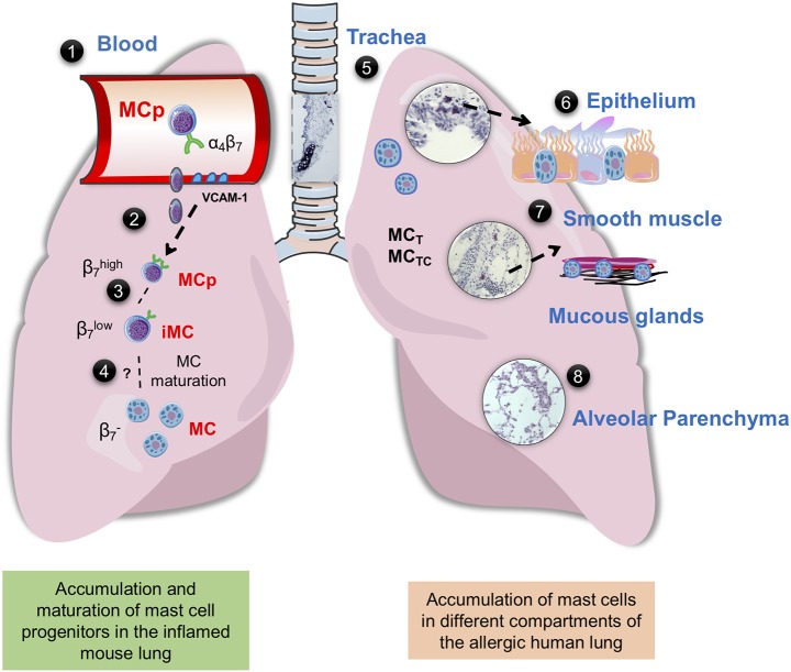
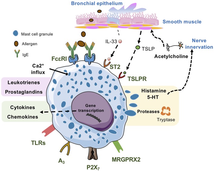
Mast cells and their mediators have been implicated in the pathogenesis of asthma and allergy for decades. Allergic asthma is a complex chronic lung disease in which several different immune cells, genetic factors and environmental exposures influence the pathology. Mast cells are key players in the asthmatic response through secretion of a multitude of mediators with pro-inflammatory and airway-constrictive effects. Well-known mast cell mediators, such as histamine and bioactive lipids are responsible for many of the physiological effects observed in the acute phase of allergic reactions. The accumulation of mast cells at particular sites of the allergic lung is likely relevant to the asthma phenotype, severity and progression. Mast cells located in different compartments in the lung and airways have different characteristics and express different mediators. According to in vivo experiments in mice, lung mast cells develop from mast cell progenitors induced by inflammatory stimuli to migrate to the airways. Human mast cell progenitors have been identified in the blood circulation. A high frequency of circulating human mast cell progenitors may reflect ongoing pathological changes in the allergic lung. In allergic asthma, mast cells become activated mainly via IgE-mediated crosslinking of the high affinity receptor for IgE (FcεRI) with allergens. However, mast cells can also be activated by numerous other stimuli e.g. toll-like receptors and MAS-related G protein-coupled receptor X2. In this review, we summarize research with implications on the role and development of mast cells and their progenitors in allergic asthma and cover selected activation pathways and mast cell mediators that have been implicated in the pathogenesis. The review places an emphasis on describing mechanisms identified using in vivo mouse models and data obtained by analysis of clinical samples.

Keywords: allergic asthma; mast cell; mast cell activation; mast cell development; mast cell progenitors.


Figures

Figure 1
Figure 1
Mast cells in mouse and human lung. (1) Integrin-β7+ mast cell progenitors (MCps) are found in mouse and human peripheral blood (14, 23). (2) In mice with acute allergic airway inflammation, MCps are recruited to the lungs in a process dependent on α4β1 and α4β7 integrins on the MCp and on VCAM-1 expressed in the endothelium (36). (3) After the acute phase, three mast cell (MC) populations can be identified by flow cytometry, MCps expressing high levels of integrin β7, immature/induced MCs (iMCs) expressing intermediate levels of integrin β7, and mature MCs (37, 38). (4) The iMC gradually loses the expression of integrin β7 and mature, thereby expanding the resident lung MC population. (5) In the mouse trachea and in the proximal bronchi of unprovoked mouse airways, MCs express the MC proteases mMCP-1 and-2, mMCP-4,-5-6, and-7 and CPA3, while the MCs induced by allergic airway inflammation located in the bronchovascular bundles of the lung and the epithelial lining of the large bronchi express mMCP-1,-2 and -6,-7 (29). In the human lung MCT and MCTC coexist, MCT is more frequently found in the bronchi, bronchioles and alveolar parenchyma, whereas MCTC dominates in pulmonary vessels and pleura (35) (6) In the human bronchi, patients with “Th2-high” asthma have an increased number of intraepithelial MCs (39). Genetic analyses suggest that the MCs in this location mainly express tryptase and CPA3 (–41). (7) The number of MCs are increased in the airway smooth muscle of asthma patients (42, 43). In diseased asthma patients, there are an increased number of mast cells in the distal airway, especially in the smooth muscle and mucous glands (44). (8) Uncontrolled atopic asthmatics have a high number of mast cells in the alveolar parenchyma (45). The histology pictures shown are from hematoxylin/eosin-stained lung sections of house dust mite-sensitized wild-type BALB/c mice obtained from our unpublished experiments.
Figure 2
Figure 2
Lung mast cells can be activated by many kinds of stimuli. IgE/antigen-mediated activation of FcεRI triggers exocytosis of granular compounds, a rapid generation and release of lipid mediators such as leukotrienes and prostaglandins and the synthesis and release of cytokines and chemokines, which occurs hours after the activation event. Mast cell activation via MRGPRX2, the adenosine A3 receptor, and the ATP receptor P2X7 also triggers release of all three types of mast cell-derived compounds. Activation via TLRs generally triggers synthesis and release of cytokines and chemokines, and some TLRs may also trigger release of lipid mediators. However, this mode of mast cell activation does not induce degranulation. IL-33, which activates the ST2 receptor complex potentiates IgE/antigen-mediated degranulation in human mast cells but not in mouse mast cells. Alone, IL-33 triggers synthesis and release of cytokines and chemokines. In mice, TSLP acting via TSLPR promotes mast cell development, whereas in human mast cells TSLP potentiates IL-33-stimulated secretion of type 2 cytokines and chemokines.

Similar articles

Cited by

References

    1. Rodewald HR, Dessing M, Dvorak AM, Galli SJ. Identification of a committed precursor for the mast cell lineage. Science. (1996) 271:818–22. 10.1126/science.271.5250.818 - DOI - PubMed
    1. Sonoda T, Hayashi C, Kitamura Y. Presence of mast cell precursors in the yolk sac of mice. Dev Biol. (1983) 97:89. 10.1016/0012-1606(83)90066-0 - DOI - PubMed
    1. Gentek R, Ghigo C, Hoeffel G, Bulle MJ, Msallam R, Gautier G, et al. . Hemogenic endothelial fate mapping reveals dual developmental origin of mast cells. Immunity. (2018) 48:1160–71.e5. 10.1016/j.immuni.2018.04.025 - DOI - PubMed
    1. Li Z, Liu S, Xu J, Zhang X, Han D, Liu J, et al. . Adult connective tissue-resident mast cells originate from late erythro-myeloid progenitors. Immunity. (2018) 49:640–53 e5. 10.1016/j.immuni.2018.09.023 - DOI - PubMed
    1. Jamur MC, Grodzki AC, Berenstein EH, Hamawy MM, Siraganian RP, Oliver C. Identification and characterization of undifferentiated mast cells in mouse bone marrow. Blood. (2005) 105:4282–9. 10.1182/blood-2004-02-0756 - DOI - PubMed

Publication types