

A β2-Integrin/MRTF-A/SRF Pathway Regulates Dendritic Cell Gene Expression, Adhesion, and Traction Force Generation
- PMID: 31191527
- PMCID: PMC6546827
- DOI: 10.3389/fimmu.2019.01138
A β2-Integrin/MRTF-A/SRF Pathway Regulates Dendritic Cell Gene Expression, Adhesion, and Traction Force Generation
Abstract
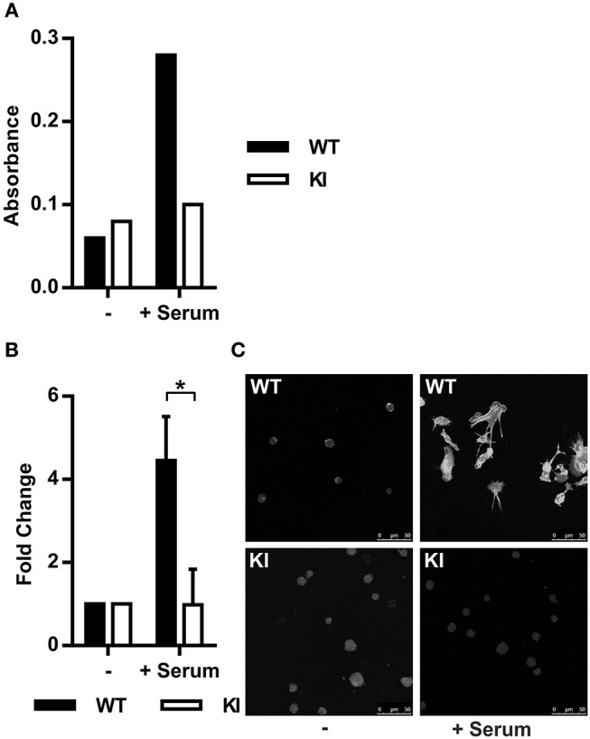
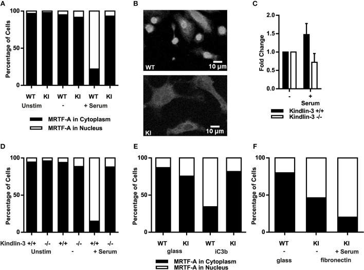
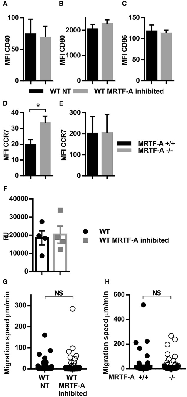
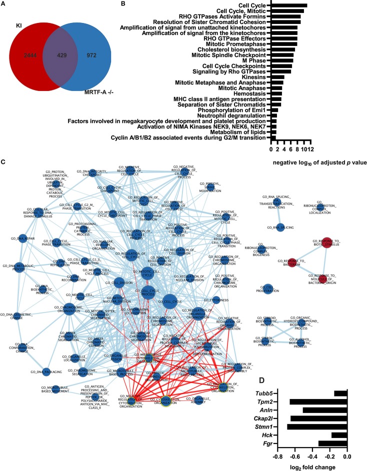
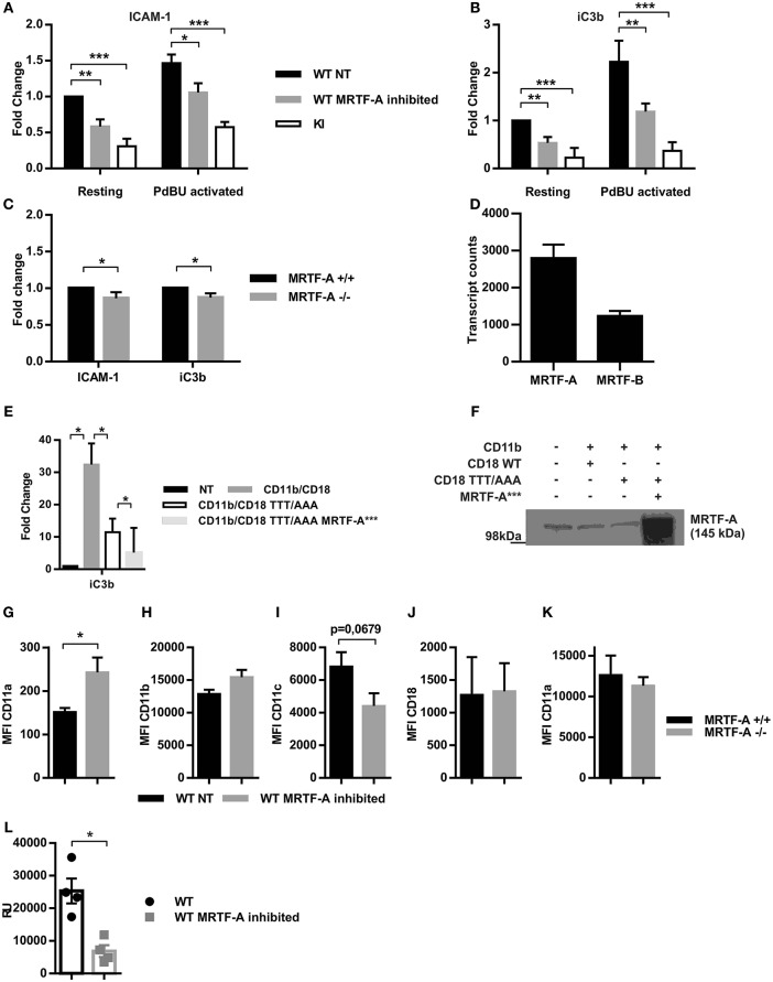
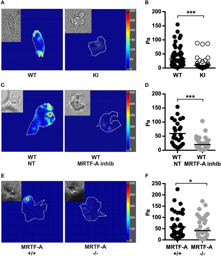
β2-integrins are essential for immune system function because they mediate immune cell adhesion and signaling. Consequently, a loss of β2-integrin expression or function causes the immunodeficiency disorders, Leukocyte Adhesion Deficiency (LAD) type I and III. LAD-III is caused by mutations in an important integrin regulator, kindlin-3, but exactly how kindlin-3 regulates leukocyte adhesion has remained incompletely understood. Here we demonstrate that mutation of the kindlin-3 binding site in the β2-integrin (TTT/AAA-β2-integrin knock-in mouse/KI) abolishes activation of the actin-regulated myocardin related transcription factor A/serum response factor (MRTF-A/SRF) signaling pathway in dendritic cells and MRTF-A/SRF-dependent gene expression. We show that Ras homolog gene family, member A (RhoA) activation and filamentous-actin (F-actin) polymerization is abolished in murine TTT/AAA-β2-integrin KI dendritic cells, which leads to a failure of MRTF-A to localize to the cell nucleus to coactivate genes together with SRF. In addition, we show that dendritic cell gene expression, adhesion and integrin-mediated traction forces on ligand coated surfaces is dependent on the MRTF-A/SRF signaling pathway. The participation of β2-integrin and kindlin-3-mediated cell adhesion in the regulation of the ubiquitous MRTF-A/SRF signaling pathway in immune cells may help explain the role of β2-integrin and kindlin-3 in integrin-mediated gene regulation and immune system function.
Keywords: LAD-III; MKL-1; MRTF-A; SRF; adhesion; dendritic cells; traction force.
Figures
Similar articles
-
The β2 integrin-kindlin-3 interaction is essential for T-cell homing but dispensable for T-cell activation in vivo.Blood. 2013 Aug 22;122(8):1428-36. doi: 10.1182/blood-2013-02-484998. Epub 2013 Jul 3. Blood. 2013. PMID: 23823319 Free PMC article.
-
Myocardin-related transcription factor A (MRTF-A) activity-dependent cell adhesion is correlated to focal adhesion kinase (FAK) activity.Oncotarget. 2016 Nov 1;7(44):72113-72130. doi: 10.18632/oncotarget.12350. Oncotarget. 2016. PMID: 27708220 Free PMC article.
-
Optimal T Cell Activation and B Cell Antibody Responses In Vivo Require the Interaction between Leukocyte Function-Associated Antigen-1 and Kindlin-3.J Immunol. 2015 Jul 1;195(1):105-15. doi: 10.4049/jimmunol.1402741. Epub 2015 May 18. J Immunol. 2015. PMID: 25987740
-
SRF'ing and SAP'ing - the role of MRTF proteins in cell migration.J Cell Sci. 2018 Oct 11;131(19):jcs218222. doi: 10.1242/jcs.218222. J Cell Sci. 2018. PMID: 30309957 Free PMC article. Review.
-
The regulatory role of serum response factor pathway in neutrophil inflammatory response.Curr Opin Hematol. 2015 Jan;22(1):67-73. doi: 10.1097/MOH.0000000000000099. Curr Opin Hematol. 2015. PMID: 25402621 Free PMC article. Review.
Cited by
-
IL-6 contributes to metastatic switch via the differentiation of monocytic-dendritic progenitors into prometastatic immune cells.J Immunother Cancer. 2021 Jun;9(6):e002856. doi: 10.1136/jitc-2021-002856. J Immunother Cancer. 2021. PMID: 34140316 Free PMC article.
-
Tuning immunity through tissue mechanotransduction.Nat Rev Immunol. 2023 Mar;23(3):174-188. doi: 10.1038/s41577-022-00761-w. Epub 2022 Aug 16. Nat Rev Immunol. 2023. PMID: 35974148 Free PMC article. Review.
-
Molecular Mechanisms of Leukocyte Migration and Its Potential Targeting-Lessons Learned From MKL1/SRF-Related Primary Immunodeficiency Diseases.Front Immunol. 2021 Feb 22;12:615477. doi: 10.3389/fimmu.2021.615477. eCollection 2021. Front Immunol. 2021. PMID: 33692789 Free PMC article. Review.
-
Actin dynamics in protein homeostasis.Biosci Rep. 2022 Sep 30;42(9):BSR20210848. doi: 10.1042/BSR20210848. Biosci Rep. 2022. PMID: 36043949 Free PMC article. Review.
-
WAVE2 Is a Vital Regulator in Myogenic Differentiation of Progenitor Cells through the Mechanosensitive MRTFA-SRF Axis.Cells. 2023 Dec 20;13(1):9. doi: 10.3390/cells13010009. Cells. 2023. PMID: 38201213 Free PMC article.
References
-
- MacPherson M, Lek H, Morrison VL, Fagerholm SC. Leukocyte beta2-integrins; genes and disease. J Genet Syndr Gene Ther. (2013) 4:154 10.4172/2157-7412.1000154 - DOI
Publication types
MeSH terms
Substances
LinkOut - more resources
Full Text Sources
Molecular Biology Databases
Miscellaneous