. 2019 May 2:2019:6904638.
doi: 10.1155/2019/6904638. eCollection 2019.

Mesenchymal Stem Cells Derived and Cultured from Glioblastoma Multiforme Increase Tregs, Downregulate Th17, and Induce the Tolerogenic Phenotype of Monocyte-Derived Cells

Affiliations

Mesenchymal Stem Cells Derived and Cultured from Glioblastoma Multiforme Increase Tregs, Downregulate Th17, and Induce the Tolerogenic Phenotype of Monocyte-Derived Cells

Kalina Tumangelova-Yuzeir et al. Stem Cells Int. .

Abstract

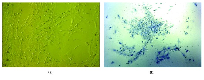
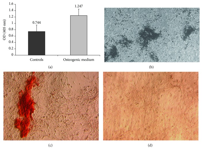
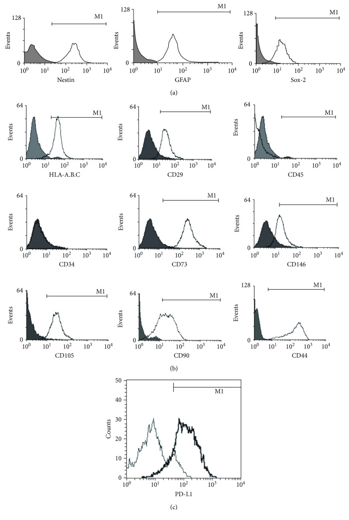
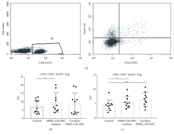
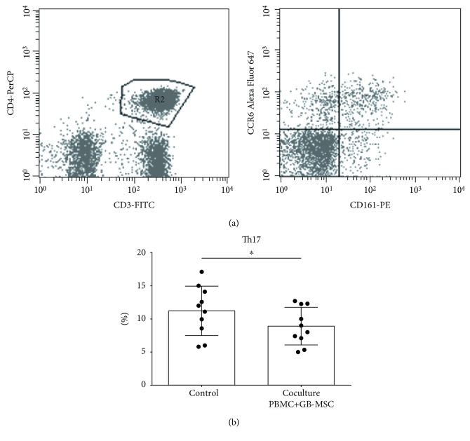
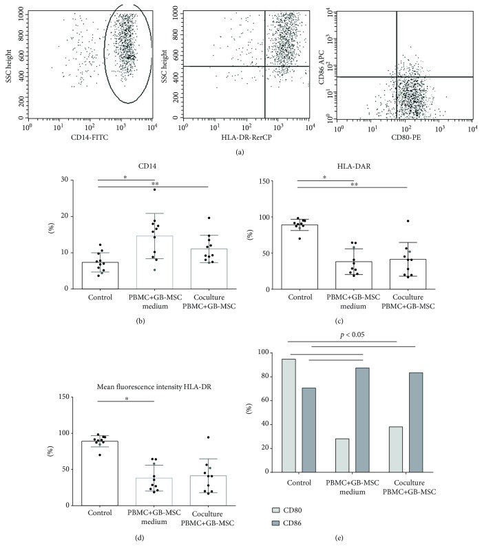
Mesenchymal stem cells (MSCs) possess immunosuppressive properties and have been described in the tumor microenvironment of glioblastoma multiforme (GBM). This manuscript has two major topics-first, to describe isolated and cultured MSCs derived from GBM (GB-MSCs) and second, to examine their in vitro immunosuppressive capacity. Our results display cells with morphology and phenotype, clonogenic ability, and osteogenic potential, typical for MSCs. Furthermore, the cultured cells show intracellular expression of the neural markers Nestin and GFAP. They express PD-L1 and secrete TGFβ, CCL-2, PGE2, IL-6, and sVEGF. Coculturing of GB-MSCs with PBMCs isolated from healthy donors results in a decreased percentage of Th17 lymphocytes and an increased percentage of Tregs. Regarding the impact of GB-MSCs on monocytes, we establish an augmented expression of CD14 and CD86 along with diminished expression of HLA-DR and CD80, which is associated with tolerogenic phenotype monocyte-derived cells. In conclusion, our results describe in detail GBM-derived and cultured cells that meet the criteria for MSCs but at the same time express Nestin and GFAP. GB-MSCs express and secrete suppressive molecules, influencing in vitro T cells and monocytes, and are probably another factor involved in the immune suppression exerted by GBM.


Figures

Figure 1
Figure 1
Morphology and clonogenicity of GB-MSCs. GB-MSCs demonstrate adherent growth and fibroblast-like morphology typical for “classical” MSCs (a). GB-MSCs demonstrated clonogenic capacity by colony formation. Crystal violet staining (b).
Figure 2
Figure 2
Differentiation of GB-MSCs in osteogenic direction. Proven by quantification of alkaline phosphatase activity (a), von Kossa (b), and alizarin red staining (c). Control cells cultured in noninductive media (d).
Figure 3
Figure 3
Marker expression by GB-MSCs. GB-MSCs have positive expression for Nestin (mean 97.1%), Sox-2 (mean 79.3%), and GFAP (mean 71.3%) (a). GB-MSCs express characteristics typical for “classical” MSC markers. More than 90% of the cells expressed CD73, CD90, CD105, CD29, CD44, CD146, and HLA-A, B, C but not CD34 and CD45 (b). GB-MSCs express PD-L1. On average, 73.2% of the cells in the cell cultures expressed this marker (c).
Figure 4
Figure 4
GB-MSCs lead to increased percentage of Tregs. Representative dot plot analysis of FoxP3+ T helper cells in the pool of PBMCs (a). Distribution of CD4+CD25-FoxP3+ T helper cells in the pool of the control PBMCs, compared to those cultured with the supernatants from GB-MSCs (p = 0.007) and cocultured with GB-MSCs (b). Distribution of CD4+CD25+FoxP3+Treg cells in the pool of the control PBMCs compared to those cultured with the supernatants from GB-MSCs and cocultured with GB-MSCs; p = 0.028 and p = 0.009, respectively (c).
Figure 5
Figure 5
GB-MSCs downregulate the percentage of Th17 lymphocytes. Representative dot plot analysis of Th17 cells in the pool of PBMCs (a). The percentage of Th17 cells in control PBMCs, compared to those cocultured with GB-MSCs, p = 0.028 (b).
Figure 6
Figure 6
GB-MSCs induce tolerogenic phenotype monocyte-derived cells. Representative dot plot analysis of monocyte-derived cell detection in the pool of PBMCs (a). Comparison of the percentage of CD14-positive monocyte-derived cells in the composition of control PBMCs, PBMCs cultured with the supernatant from GB-MSCs (p = 0.005), and PBMCs cocultured with GB-MSCs (p = 0.007) (b). Comparison of the percentage of monocyte-derived cells expressing HLA-DR in monocyte-derived cells in the pool of control PBMCs and PBMCs cultured with supernatants of GB-MSCs (p = 0.005) and PBMCs cocultured with GB-MSCs (p = 0.005) (c). Comparison of the HLA-DR mean fluorescent intensity (MFI) in monocyte-derived cells in the pool of control PBMCs and PBMCs cultured with supernatants of GB-MSCs (p = 0.011) and PBMCs cocultured with GB-MSCs (d). Comparison of the percentage of CD80- and CD86-expressing monocyte-derived cells in the composition of control PBMCs and PBMCs cultured with the supernatant from GB-MSCs (p = 0.028 and p = 0.028, respectively) and PBMCs cocultured with GB-MSCs (p = 0.028 and p = 0.028, respectively) (e).

Similar articles

Cited by

References

    1. Louis D., Ellison D. Greenfield’s Neuropathology. 8th Edition. Hodder Arnold, Seth Love; 2008.
    1. Wen P., Kesari S. Malignant gliomas in adults. The New England Journal of Medicine. 2008;359(5):492–507. doi: 10.1056/NEJMra0708126. - DOI - PubMed
    1. Modrek A. S., Bayin N. S., Placantonakis D. G. Brain stem cells as the cell of origin in glioma. World Journal of Stem Cells. 2014;6(1):43–52. doi: 10.4252/wjsc.v6.i1.43. - DOI - PMC - PubMed
    1. Molina E. S., Pillat M. M., Moura-Neto V., Lah T. T., Ulrich H. Glioblastoma stem-like cells: approaches for isolation and characterization. Journal of Cancer Stem Cell Research. 2014;2, article e1007 doi: 10.14343/jcscr.2014.2e1007. - DOI
    1. Bayin N. S., Modrek A. S., Placantonakis D. G. Glioblastoma stem cells: molecular characteristics and therapeutic implications. World Journal of Stem Cells. 2014;6(2):230–238. doi: 10.4252/wjsc.v6.i2.230. - DOI - PMC - PubMed