. 2019 May 2:2019:9545261.
doi: 10.1155/2019/9545261. eCollection 2019.

Identification of Macrophage Genotype and Key Biological Pathways in Circulating Angiogenic Cell Transcriptome

Affiliations

Identification of Macrophage Genotype and Key Biological Pathways in Circulating Angiogenic Cell Transcriptome

Bert R Everaert et al. Stem Cells Int. .

Abstract

Background: Circulating angiogenic cells (CAC) have been identified as important regulators of vascular biology. However, there is still considerable debate about the genotype and function of CAC.

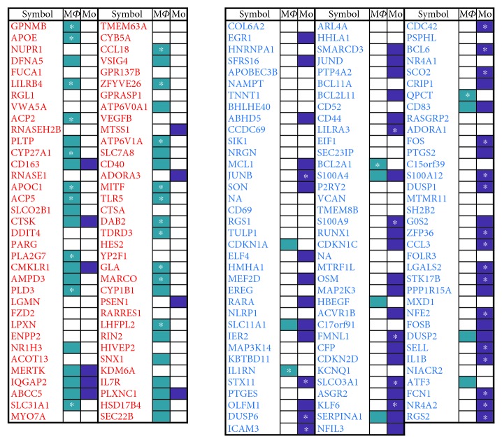
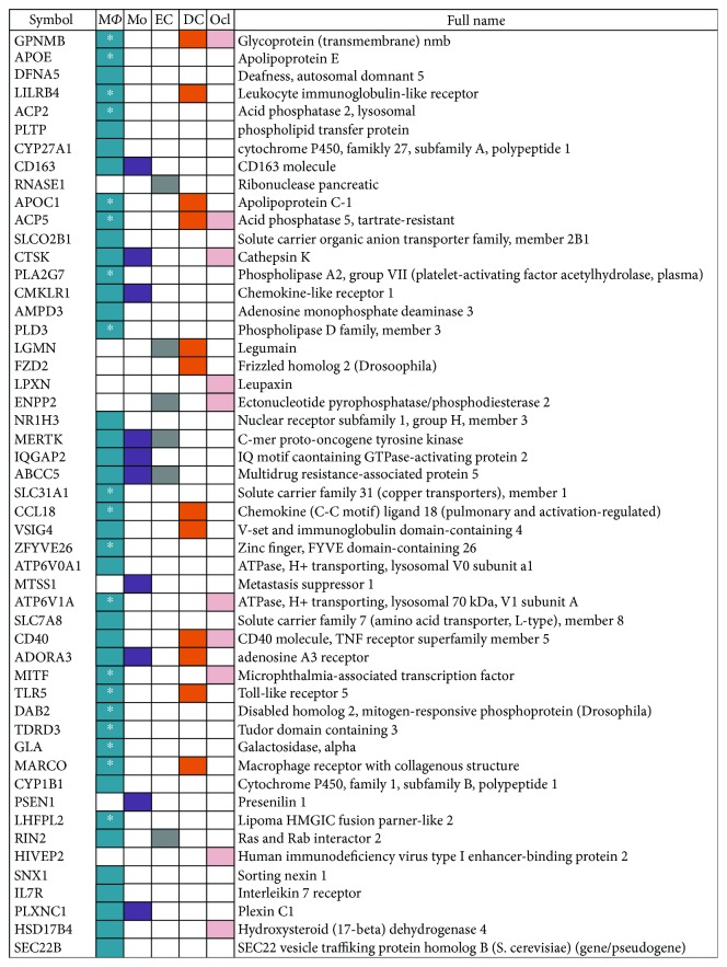
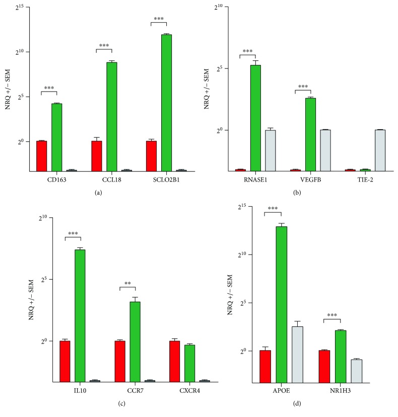
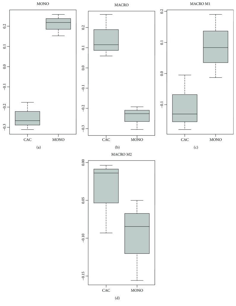
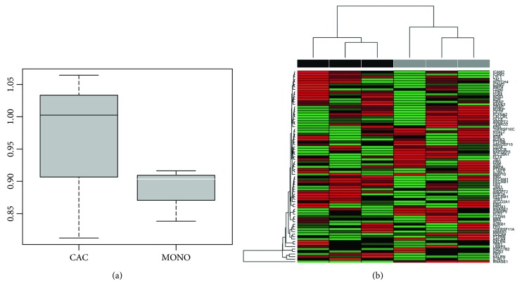
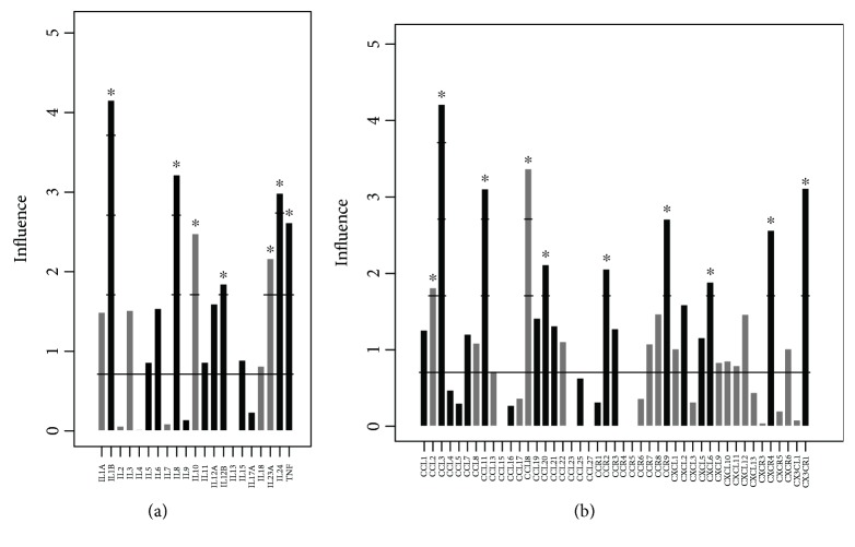
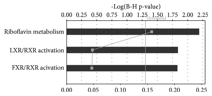
Methods and results: Data from publicly available gene expression data sets were used to analyse the transcriptome of in vitro cultured CAC (CACiv). Genes and pathways of interest were further evaluated using qPCR comparing CACiv versus CD14+ monocytic cells. The CACiv transcriptome strongly related to tissue macrophages, and more specifically to regulatory M2c macrophages. The cytokine expression profile of CACiv was predominantly immune modulatory and resembled the cytokine expression of tumor-associated macrophages (TAM). Pathway analysis revealed previously unrecognized biological processes in CACiv, such as riboflavin metabolism and liver X receptor (LXR)/retinoid X receptor (RXR) and farnesoid X receptor (FXR)/retinoid X receptor (RXR) pathways. Analysis of endothelial-specific genes did not show evidence for endothelial transdifferentiation.

Conclusions: CACiv are genotypically similar to regulatory M2c macrophages and lack signs of endothelial differentiation.


Figures

Figure 1
Figure 1
CACiv gene signature. 70 genes were significantly upregulated versus 107 genes were significantly downregulated in CACiv versus CD14+ monocytic cells. The upregulated (red) and downregulated (blue) genes express a close lineage relationship with macrophages and monocytes, respectively. We highlighted those genes of the CACiv signature that are specific for either macrophages or monocytes. : very specific. Mϕ: macrophage; Mo: monocyte.
Figure 2
Figure 2
Expression of CACiv signature related genes in myeloid cells. To demonstrate the lineage relationships between CACiv and other cell types, we highlighted those genes of the CACiv signature that were found to be significantly upregulated and also specific for a particular cell type. : very specific for macrophages. Mϕ: macrophage; Mo: monocyte; EC: endothelial cell; DC: dendritic cell; Ocl: osteoclast.
Figure 3
Figure 3
Gene expression analysis. Gene expression of (a) M2c macrophage-related, (b) endothelial, (c) cytokine/cytokine receptor and (d) cholesterol transporter pathway genes in CACiv (green) vs. CD14+ monocytes (red) vs. human umbilical vein endothelial cells (HUVEC) (grey). Only the relevant statistical significance between CACiv and CD14+ monocytes is depicted. NRQ: normalized relative quantity. ∗∗p < 0.01; ∗∗∗p < 0.001.
Figure 4
Figure 4
CACiv differentiate towards a predominanty M2 macrophage genotype. Boxplots comparing the relative expression of the genes in (a) the monocyte-specific gene signature (MONO), (b) the macrophage-specific gene signature (MACRO), (c) the LPS- and IFN-γ-stimulated M1 macrophage-specific gene signature (MACRO M1), and (d) the IL4-stimulated M2 macrophage-specific gene signature (MACRO M2), between CACiv and CD14+ monocytic cells (MONO). Positive values signify a stronger degree of similarity of the genetic expression of CACiv or CD14+ monocytic cells to the specified cell type.
Figure 5
Figure 5
Analysis of EC-associated genes in the CACiv profile. (a) Boxplot comparing the relative expression of an endothelial gene set between CACiv and CD14+ monocytic cells. There is no significant difference in similarity of the genetic expression of CACiv or CD14+ monocytic cells to an EC-specific gene signature. (b) UHC analysis highlighting the relative expression of EC-associated genes in CACiv (grey) and monocytes (black). RNASE1 is indicated as most differentially expressed in CACiv compared to CD14+ monocytic cells. We observed a lack of consistency in EC-associated gene expression between different CACiv culture samples. Red: upregulation; green: downregulation.
Figure 6
Figure 6
CACiv -related cytokine and chemokine/chemokine receptor expression profile. Gene expression plot depicting (a) cytokines or (b) chemokines and chemokine receptors that were differentially overexpressed in CACiv (grey) vs. monocytes (black). The influence (y-axis) represents the number of standard deviations (SD) the gene expression of each gene exceeds the null hypothesis that there would be no difference between both groups (z-score). Genes with an influence of ≥1.96 show a statistically significant differential gene expression (p < 0.05). p < 0.05.
Figure 7
Figure 7
Summary of IPA analysis. The main upregulated canonical pathways are shown. Riboflavin metabolism and LXR/RXR and FXR/RXR activation remain statistically significant even after correction for multiple comparisons (Threshold indicates false discovery rate corrected p-value of 0.05). Bottom axis depicts the ratio of the number of genes upregulated in de CACiv signature divided by the total number of genes involved in a given pathway (squares).

References

    1. Asahara T., Murohara T., Sullivan A., et al. Isolation of putative progenitor endothelial cells for angiogenesis. Science. 1997;275(5302):964–966. doi: 10.1126/science.275.5302.964. - DOI - PubMed
    1. Rehman J., Li J., Orschell C. M., March K. L. Peripheral blood "endothelial progenitor cells" are derived from monocyte/macrophages and secrete angiogenic growth factors. Circulation. 2003;107(8):1164–1169. doi: 10.1161/01.CIR.0000058702.69484.A0. - DOI - PubMed
    1. Hur J., Yoon C. H., Kim H. S., et al. Characterization of two types of endothelial progenitor cells and their different contributions to neovasculogenesis. Arteriosclerosis, Thrombosis, and Vascular Biology. 2004;24(2):288–293. doi: 10.1161/01.ATV.0000114236.77009.06. - DOI - PubMed
    1. Urbich C., de Souza A. I., Rossig L., et al. Proteomic characterization of human early pro-angiogenic cells. Journal of Molecular and Cellular Cardiology. 2011;50(2):333–336. doi: 10.1016/j.yjmcc.2010.11.022. - DOI - PubMed
    1. Aicher A., Heeschen C., Mildner-Rihm C., et al. Essential role of endothelial nitric oxide synthase for mobilization of stem and progenitor cells. Nature Medicine. 2003;9(11):1370–1376. doi: 10.1038/nm948. - DOI - PubMed