

Traditional Herbal Formula Taeeumjowi-Tang (TJ001) Inhibits p53-Mutant Prostate Cancer Cells Growth by Activating AMPK-Dependent Pathway
- PMID: 31191706
- PMCID: PMC6525874
- DOI: 10.1155/2019/2460353
Traditional Herbal Formula Taeeumjowi-Tang (TJ001) Inhibits p53-Mutant Prostate Cancer Cells Growth by Activating AMPK-Dependent Pathway
Abstract
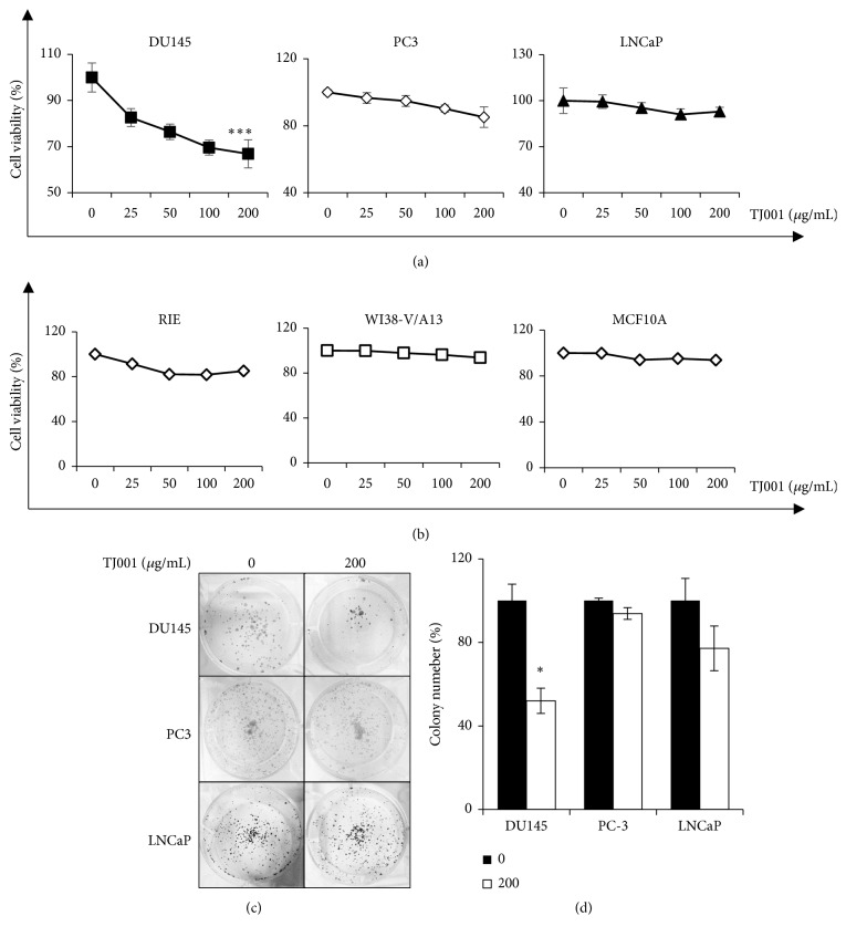
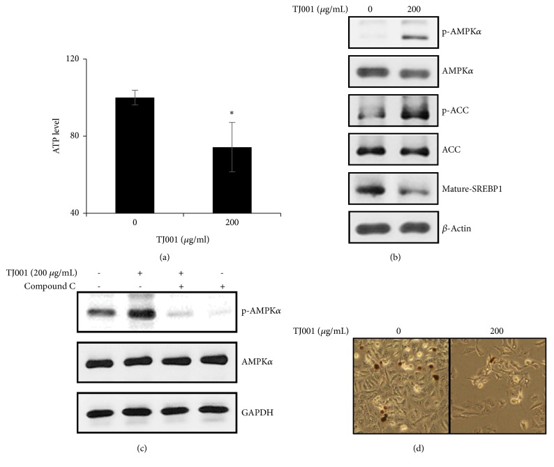
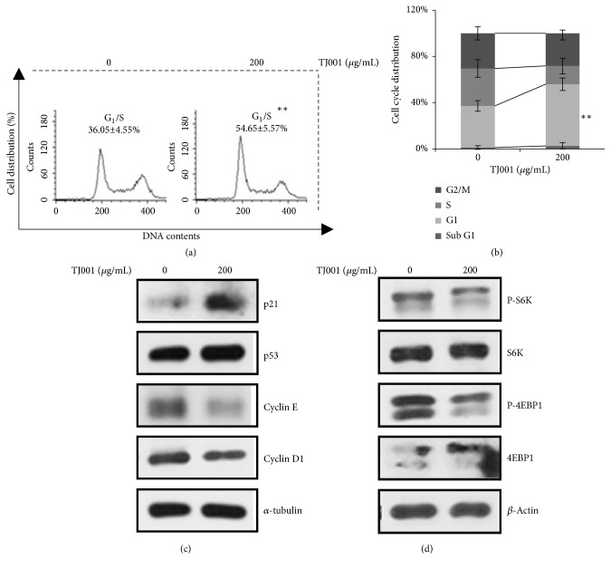
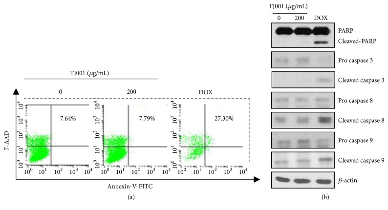
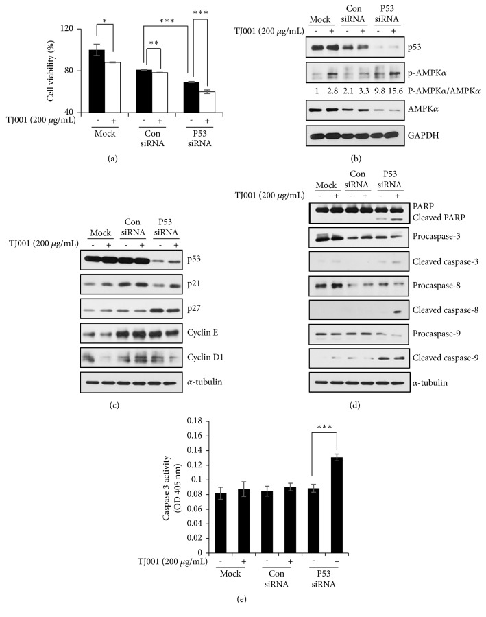
Dysregulated lipid metabolism is a prominent feature of prostate cancers (PCas); several enzymes involved in lipid accumulation are highly expressed. Here, we elucidated efficacy of TJ001, a traditional herbal decoction, in inhibiting de novo lipogenesis. TJ001 had significant cytotoxicity against DU145 but not PC3 and LNCaP cells and, similarly, TJ001 markedly AMPK phosphorylation only in DU145 cells. This was accompanied by the downregulation of phosphorylated-acetyl coenzyme A carboxylase (ACC) expression and sterol regulatory element-binding protein 1 (SREBP1) proteolytic cleavage, thereby inhibiting its role as a transcription factor to induce lipid biosynthesis. When Oil Red O staining was performed, it is reflected in the reduction of lipid droplets (LDs). TJ001 also induced G1/S cell cycle arrest via a cell cycle inhibitor (CKI) p21WAF1/CIP1 upregulation. Although p53 proteins remained unchanged, both cyclin E and cyclin D1 were decreased. Moreover, TJ001 suppressed the mammalian target of rapamycin (mTOR) signaling pathway. Generally, the prolonged G1/S phase arrest accompanies apoptosis, but TJ001 failed to work as a trigger apoptosis in DU145 cells. We showed that mutant p53 proteins were required for the survival of DU145 cells. In presence of TJ001, inhibition of endogenous mutant p53 by RNAi led to cell viability reduction and induction of the p-AMPK/AMPK ratio. In addition, it induced apoptotic cell death in DU145 cells. At the cellular level, induction of PARP, caspase-3, and caspase-9 cleavages was observed, and caspase-3 activity was increased in the p53 knockdown cells treated with TJ001. Taken together, we demonstrated that TJ001 inhibited cell growth in DU145 prostate cancer cells as indicated by blocking lipogenesis and induction in G1/S cell cycle arrest. In addition, we may provide an evidence that mutant p53 protein has potential role as an oncogenic action in DU145 cells. Collectively, the combination of mutant p53 targeting and TJ001 treatment resulted in decreased cell growth in DU145 cells.
Figures
Similar articles
-
Persistent p21Cip1 induction mediates G(1) cell cycle arrest by methylseleninic acid in DU145 prostate cancer cells.Curr Cancer Drug Targets. 2010 May;10(3):307-18. doi: 10.2174/156800910791190238. Curr Cancer Drug Targets. 2010. PMID: 20370687
-
LYG-202 inhibits the proliferation of human colorectal carcinoma HCT-116 cells through induction of G1/S cell cycle arrest and apoptosis via p53 and p21(WAF1/Cip1) expression.Biochem Cell Biol. 2011 Jun;89(3):287-98. doi: 10.1139/o10-162. Epub 2011 Apr 14. Biochem Cell Biol. 2011. PMID: 21491996
-
Molecular pathway for (-)-epigallocatechin-3-gallate-induced cell cycle arrest and apoptosis of human prostate carcinoma cells.Arch Biochem Biophys. 2003 Feb 1;410(1):177-85. doi: 10.1016/s0003-9861(02)00668-9. Arch Biochem Biophys. 2003. PMID: 12559991
-
A flavonoid antioxidant, silymarin, inhibits activation of erbB1 signaling and induces cyclin-dependent kinase inhibitors, G1 arrest, and anticarcinogenic effects in human prostate carcinoma DU145 cells.Cancer Res. 1998 May 1;58(9):1920-9. Cancer Res. 1998. PMID: 9581834
-
Inositol hexaphosphate inhibits growth, and induces G1 arrest and apoptotic death of prostate carcinoma DU145 cells: modulation of CDKI-CDK-cyclin and pRb-related protein-E2F complexes.Carcinogenesis. 2003 Mar;24(3):555-63. doi: 10.1093/carcin/24.3.555. Carcinogenesis. 2003. PMID: 12663518
Cited by
-
Gardenia jasminoides Enhances CDDP-Induced Apoptosis of Glioblastoma Cells via AKT/mTOR Pathway While Protecting Death of Astrocytes.Nutrients. 2020 Jan 10;12(1):196. doi: 10.3390/nu12010196. Nutrients. 2020. PMID: 31936835 Free PMC article.
-
SH005S7 Overcomes Primary and Acquired Resistance of Non-Small Cell Lung Cancer by Combined MET/EGFR/HER3 Inhibition.Biomed Res Int. 2022 Sep 15;2022:1840541. doi: 10.1155/2022/1840541. eCollection 2022. Biomed Res Int. 2022. PMID: 36158893 Free PMC article.
-
Targeting SREBP-1-Mediated Lipogenesis as Potential Strategies for Cancer.Front Oncol. 2022 Jul 14;12:952371. doi: 10.3389/fonc.2022.952371. eCollection 2022. Front Oncol. 2022. PMID: 35912181 Free PMC article. Review.
-
Pediatric Finger Warts Treated Using Taeeumjowi-tang: a case report.J Pharmacopuncture. 2024 Jun 30;27(2):172-176. doi: 10.3831/KPI.2024.27.2.172. J Pharmacopuncture. 2024. PMID: 38948305 Free PMC article.
References
-
- Ecke T. H., Schlechte H. H., Schiemenz K., et al. TP53 gene mutations in prostate cancer progression. International Journal of Cancer Research and Treatments. 2010;30(5):1579–1586. - PubMed
LinkOut - more resources
Full Text Sources
Research Materials
Miscellaneous
