Review
. 2019 Mar 21;10(5):634-646.
doi: 10.1039/c9md00126c. eCollection 2019 May 1.

The calcium-dependent lipopeptide antibiotics: structure, mechanism, & medicinal chemistry

Affiliations
Review

The calcium-dependent lipopeptide antibiotics: structure, mechanism, & medicinal chemistry

Thomas M Wood et al. Medchemcomm. .

Abstract

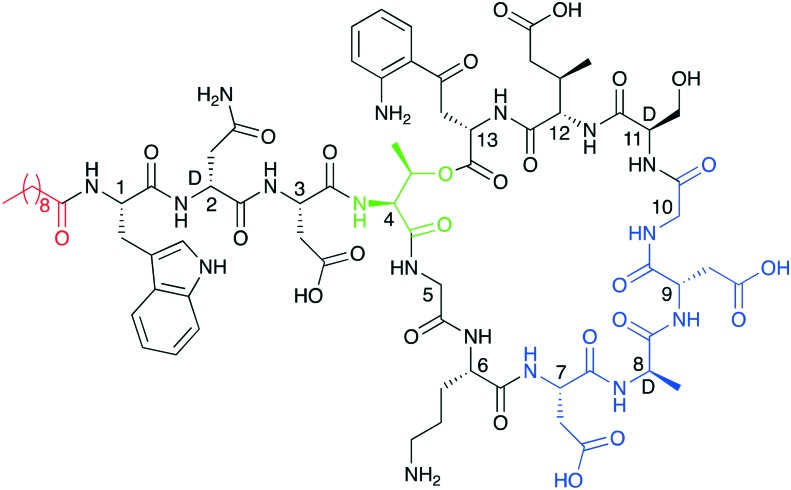
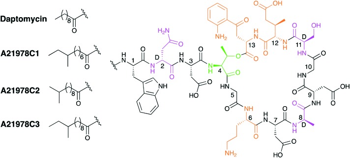
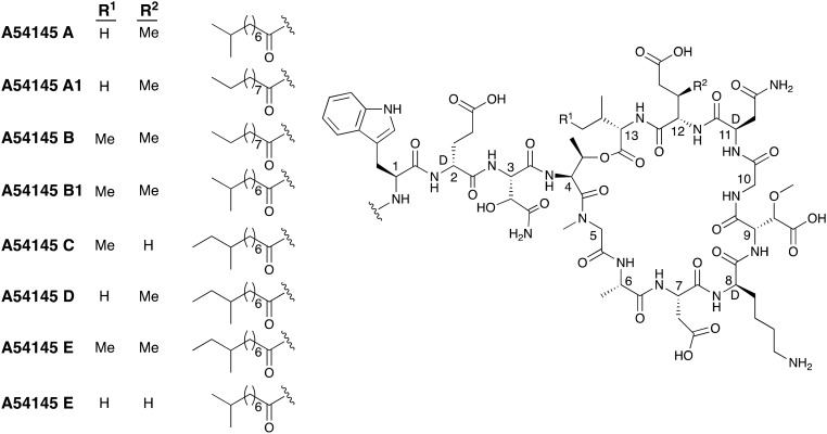
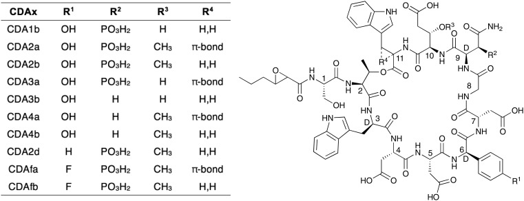
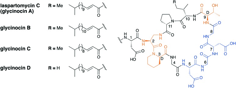
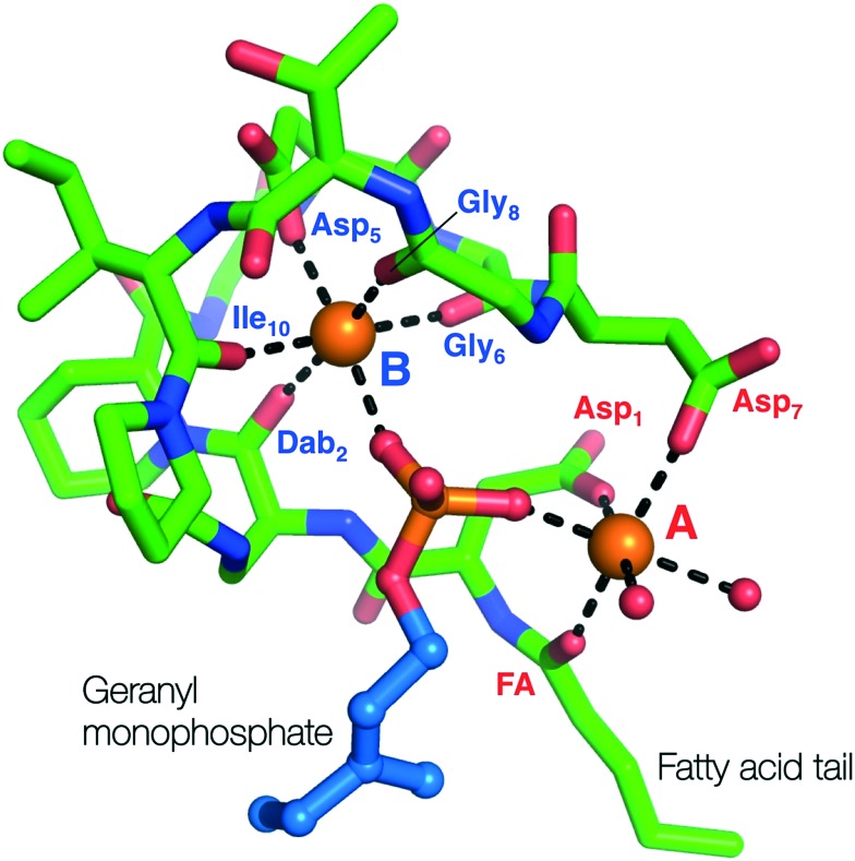
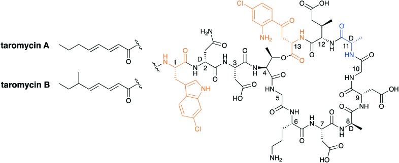
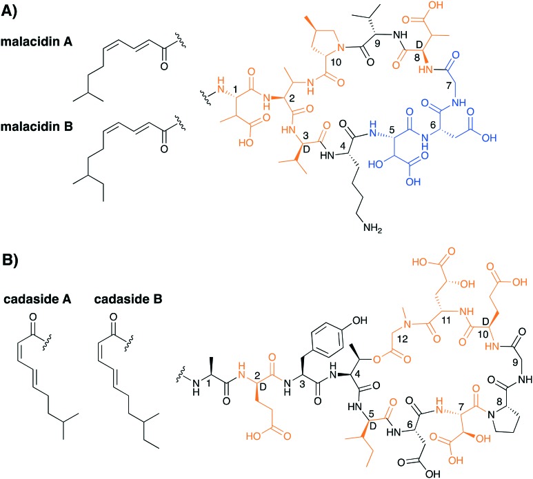
To push back the growing tide of antibacterial resistance the discovery and development of new antibiotics is a must. In recent years the calcium-dependent lipopeptide antibiotics (CDAs) have emerged as a potential source of new antibacterial agents rich in structural and mechanistic diversity. All CDAs share a common lipidated cyclic peptide motif containing amino acid side chains that specifically chelate calcium. It is only in the calcium bound state that the CDAs achieve their potent antibacterial activities. Interestingly, despite their common structural features, the mechanisms by which different CDAs target bacteria can vary dramatically. This review provides both a historic context for the CDAs while also addressing the state of the art with regards to their discovery, optimization, and antibacterial mechanisms.


Figures

Fig. 1
Fig. 1. Structure of daptomycin indicating N-terminal lipid in red and in blue the calcium binding motifs. The peptide macrocycles are formed biosynthetically via cyclization of the C-terminal residue with the side chain of Thr4 (linkages shown in green).
Fig. 2
Fig. 2. Structures of the A21978C family; the macrolactonization site is shaded in green, d-amino acids in purple and non-proteinogenic amino acids in orange.
Scheme 1
Scheme 1. SPPS approach used by Cubist team to achieve the first total synthesis of daptomycin.
Scheme 2
Scheme 2. Serine ligation route used by Li and coworkers in synthesizing daptomycin.
Scheme 3
Scheme 3. Entirely SPPS approach used by the Taylor group in the synthesis of daptomycin.
Fig. 3
Fig. 3. Structure of surotomycin. This semisynthetic analogue of daptomycin bears an unnatural lipid tail imparting potent activity against C. difficile.
Fig. 4
Fig. 4. Structures of the (A) friulimicin and (B) amphomycin families of CDA. Indicated in brackets are the carious other names that have historically been assigned to these structures.
Fig. 5
Fig. 5. Structure of the semisynthetic amphomycin analogue MX-2401. Highlighted in red is the 4-(dodecanamido)benzoic acid lipid tail and in blue the modified Dab9.
Fig. 6
Fig. 6. Structures of A54145 class of CDAs.
Fig. 7
Fig. 7. Structural diversity among the CDA class.
Fig. 8
Fig. 8. Laspartomycin C and the glycinocin A–D family. Highlighted in blue is the conserved Asp-X-Asp-Gly Ca2+ binding motif and highlighted in orange are nonproteinogenic amino acids.
Scheme 4
Scheme 4. Combined solution- and solid-phase approach used by our group in the first total synthesis of laspartomycin C.
Fig. 9
Fig. 9. Crystal structure of laspartomycin C (green stick representation) bound to two bound Ca2+ ions (orange spheres) and the geranyl monophosphate ligand coordinated by the Ca2+ ions. The two Ca2+ ions are labelled A (red) and B (blue) with the interacting parts of laspartomycin C responsible for the chelation of the Ca2+ ions also indicated.
Fig. 10
Fig. 10. Taromycin A and B. Highlighted in orange are the chlorinated amino acids 6-chloro-tryptophan and 4-chloro-kynurenine. The taromycin family has a d-alanine in place of the d-serine found in daptomycin (highlighted in blue).
Fig. 11
Fig. 11. Structures of A) the malacidins and B) the cadasides. Highlighted in orange are the nonproteinogenic amino acids and highlighted in blue is the presumed Ca2+ binding motif for the malacidins.

Similar articles

Cited by

References

    1. O'Neill J., Antimicrobial Resistance: Tackling a Crisis for the Health and Wealth of Nations, 2014, http://www.jpiamr.eu/wp-content/uploads/2014/12/AMR-Review-Paper-Tacklin....
    1. CDCD, US Dep. Heal. Hum. Serv.
    1. Waterman P. E., McGann P., Snesrud E., Clifford R. J., Kwak Y. I., Munoz-Urbizo I. P., Tabora-Castellanos J., Milillo M., Preston L., Aviles R., Sutter D. E., Lesho E. P. Antimicrob. Agents Chemother. 2013;57:4584–4586. - PMC - PubMed
    1. Tehrani K. H. M. E., Martin N. I. Med. Chem. Commun. 2018;9:1439–1456. - PMC - PubMed
    1. Wang Y., Zhang R., Li J., Wu Z., Yin W., Schwarz S., Tyrrell J. M., Zheng Y., Wang S., Shen Z., Liu Z., Liu J., Lei L., Li M., Zhang Q., Wu C., Zhang Q., Wu Y., Walsh T. R., Shen J. Nat. Microbiol. 2017;2:1–7. - PubMed