. 2019 May 2:2019:1902915.
doi: 10.1155/2019/1902915. eCollection 2019.

Investigation of Blood Characteristics in Nonsyndromic Retinitis Pigmentosa: A Retrospective Study

Affiliations

Investigation of Blood Characteristics in Nonsyndromic Retinitis Pigmentosa: A Retrospective Study

Yijing Yang et al. J Ophthalmol. .

Abstract

Purpose: To investigate the characteristics of blood in nonsyndromic retinitis pigmentosa (RP) and reveal the pathogenesis of blood cells involved in blood stasis in RP.

Design: This is a retrospective observational study.

Methods: We collected vein blood from 101 cases of patients with nonsyndromic RP and 120 cases of normal individuals according to a single-blind study and used routine clinical examination to detect the indicators of blood. All the subjects were mainly from the central south of China. Data were analyzed statistically between the RP group and normal control.

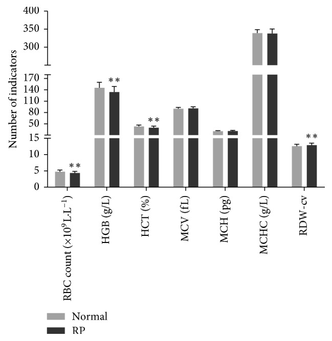
Results: The indicator of platelet distribution width (PDW) in patients with RP was higher than that in the normal group; the indicators of red blood cell (RBCs), hemoglobin (HGB), hematocrit (HCT), basophils (BASs), platelets (PLTs), and plateletcrit (PCT) in the RP group were lower than those in the normal control. The differences were statistically very significant between the RP group and normal group (p < 0.01). There were no statistical differences in the other indicators between the RP and normal group.

Conclusions: The changes in RBCs and PLTs in patients with RP implied that RP induces RBC aggregation and platelet activation, leading to blood stasis which in turn initiates more apoptosis.


Figures

Figure 1
Figure 1
Comparison of RBC indicators between the normal and RP group. All data were in accordance with the normal distribution, and two independent samples t-tests were performed for comparison. p < 0.05 was considered statistically significant; ∗∗p < 0.01 was considered statistically very significant.
Figure 2
Figure 2
Comparison of WBC indicators between the normal and RP group. Two independent samples T-tests were performed for comparing data of WBC and NEU counts. Two independent samples rank sum test was performed for other data. p < 0.05 was considered statistically significant. ∗∗p < 0.01 was considered statistically very significant.
Figure 3
Figure 3
Comparison of PLT indicators between the normal and RP group. Two independent samples T-tests were performed for comparing the data of PLT. Two independent samples rank sum test was performed for the other data. p < 0.05 was considered statistically significant. ∗∗p < 0.01 was considered statistically very significant.

Similar articles

References

    1. Hamel C. Retinitis pigmentosa. Orphanet Journal of Rare Diseases. 2006;1(1):p. 40. doi: 10.1186/1750-1172-1-40. - DOI - PMC - PubMed
    1. Hartong D. T., Berson E. L., Dryja T. P. Retinitis pigmentosa. The Lancet. 2006;368(9549):1795–1809. doi: 10.1016/s0140-6736(06)69740-7. - DOI - PubMed
    1. Falsini B., Anselmi G. M., Marangoni D., et al. Subfoveal choroidal blood flow and central retinal function in retinitis pigmentosa. Investigative Ophthalmology & Visual Science. 2011;52(2):1064–1069. doi: 10.1167/iovs.10-5964. - DOI - PubMed
    1. Beutelspacher S., Serbecic N., Hausner L., Eliash Z. B., Grinvald A., Jonas J. B. Central serous chorioretinopathy shows reduced retinal flow circulation in retinal function imaging (RFI) Acta Ophthalmologica. 2011;89(6):e479–e482. doi: 10.1111/j.1755-3768.2011.02136.x. - DOI - PubMed
    1. Murakami Y., Funatsu J., Nakatake S., et al. Relations among foveal blood flow, retinal-choroidal structure, and visual function in retinitis pigmentosa. Investigative Ophthalmology & Visual Science. 2018;59(2):1134–1143. doi: 10.1167/iovs.17-23050. - DOI - PubMed

LinkOut - more resources