. 2019 May 2:2019:4153064.
doi: 10.1155/2019/4153064. eCollection 2019.

Tectonic Keratoplasty to Restore the Bulbar Wall after Block Excision of Benign and Malignant Intraocular Tumors

Affiliations

Tectonic Keratoplasty to Restore the Bulbar Wall after Block Excision of Benign and Malignant Intraocular Tumors

Emilio Balestrazzi et al. J Ophthalmol. .

Abstract

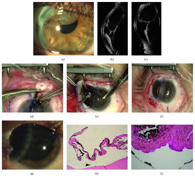
Purpose: To report the surgical treatment and follow-up of tumors of the anterior uvea and epithelial cysts of the anterior chamber in 4 patients submitted to block excision and tectonic corneal graft between 2008 and 2012.

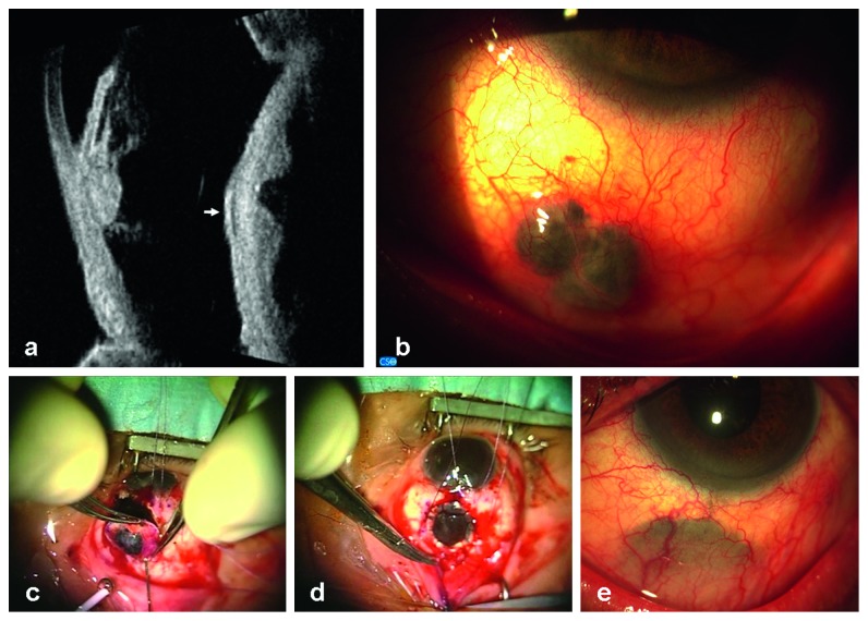
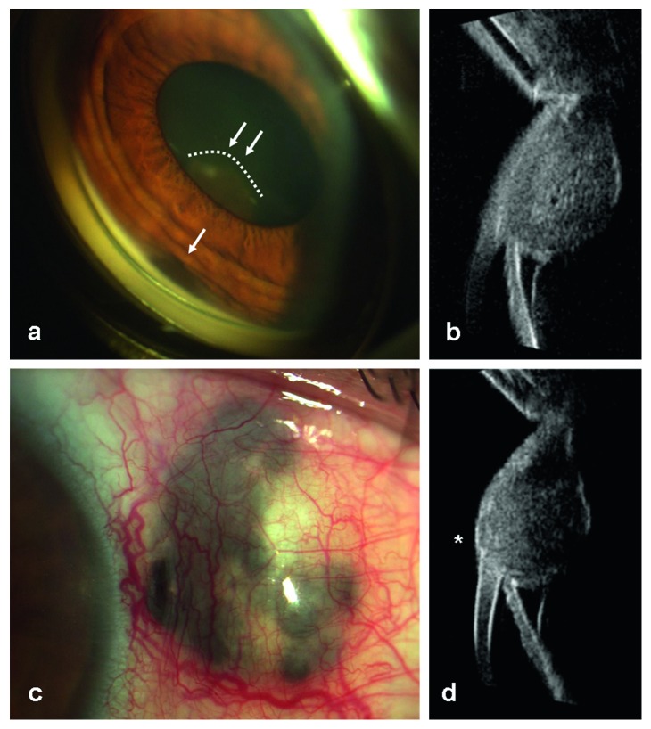
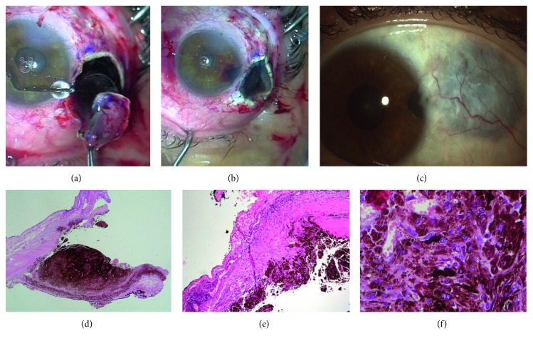
Methods: This is a retrospective, nonrandomized case series. Two patients were affected by anterior uveal malignant melanoma, and 2 patients were referred to us for large epithelial iris cysts with anterior chamber angle involvement and partial corneal failure. A simultaneous block removal of the lesion and adjacent iris, cornea (when necessary), ciliary body, and sclera was performed; the resulting defect was covered by a tectonic whole thickness corneal graft. Follow-up ranged from 2 to 7 years (mean time: 5 ± 1.6 MD).

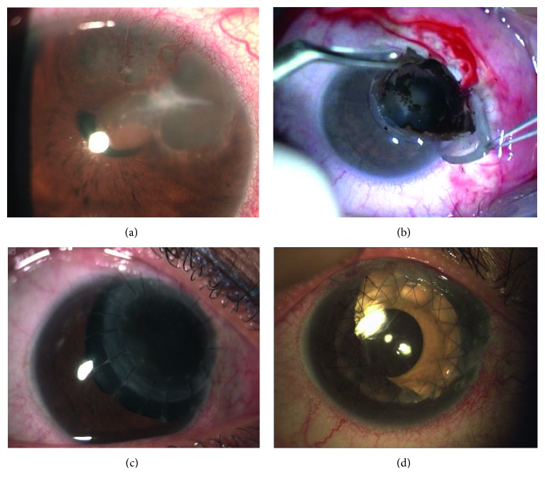
Results: Local control of malignant melanoma was observed during the follow-up, but cataract surgery was planned in both patients and pars plana vitrectomy for vitreous hemorrhage occurred in one case. No recurrence of cysts was detected. After iris cysts excision, a planned second-time surgery was necessary in one patient: optical penetrating keratoplasty, centered on the visual axis, implantation of one refractive IOL (intraocular lens) in the bag, and one cosmetic IOL in the sulcus, to restore the iris diaphragm.

Conclusions: Block excision followed by the tectonic corneal graft seems to be the treatment of choice for selected cases of epithelial cysts of the anterior chamber and anterior uvea melanomas with epibulbar extension. Further surgery, as a second step, could be required to improve functional results of this challenging technique.


Figures

Figure 1
Figure 1
Patient 1 UBM before surgery and after uveo-scleral excision en bloc. (a) Left: longitudinal scan at the 6 o'clock position (L6) shows a solid thickening of the ciliary body and the anterior choroid; the corresponding anterior chamber angle is infiltrated, the acoustic structure is heterogeneous, and the internal reflectivity is medium. Right: transversal scan at the 5:30 o'clock position (T5:30) shows the double dome-shaped morphology of the lesion at the level of the anterior choroid and the circumscribed growth into the overlying sclera, which appears hypoechoic (white arrow). (b) Patient 1: slit lamp examination presurgery. Image shows a confined pigmented nodular growth of uveal melanoma through the sclera, at about 6 mm from the limbus, under the conjunctival layer. (c) After Hanna trephination, block excision is completed with scissors until removing the entire lesion and the overlying infiltrated sclera. (d) Tectonic corneal graft covers the scleral defect and is sutured with 16 interrupted Nylon 10.0 sutures. (e) Corneal graft is well integrated into the sclera, under the conjunctiva, about 4 years after surgery.
Figure 2
Figure 2
Patient 2 before treatment. (a) Gonioscopy shows a pigmented fusiform thickening of the iris root (white arrow) and a dome-shaped pigmented mass behind the pupil foramen (dashed line and double white arrow). (b) UBM longitudinal scan at the 2:30 o'clock position (L2:30) showing a circumscribed dome-shaped mass at the level of the ciliary body, extended to the iris root, which touches the anterior surface of the lens. (c) Scleral thinning and prolapse of the uveal tissue at the limbus, one year after brachytherapy. (d) UBM L2:30 scan shows a local scleral extension of the tumor, under the conjunctival layer (white asterisk).
Figure 3
Figure 3
Patient 2 block excision of sclera and ciliary melanoma. (a) Removal of the ciliary melanoma en bloc after marking the margins and after Hanna trephination. (b) Corneal graft is sutured to the adjacent sclera; basal iridectomy. (c) Corneal graft is in situ 7 years after surgery well visible under the conjunctiva. (d) The perfect clearance of trephination on healthy scleral and ciliary body margins. (e, f) Postirradiation necrotic aspect of spindle melanoma's cells.
Figure 4
Figure 4
Patient 3 before and after surgery. (a) Large epithelial cysts of the iris involving the upper nasal quadrant of the right eye. Cysts stretch the pupil foramen and cause circumscribed corneal failure. (b) Block excision of the cysts, iris, cornea, and limbal sclera after Hanna trephination. (c) Eccentric corneal graft is clear one-year postoperatively, and sutures are still in situ; a large iris coloboma is observable. (d) Two months after penetrating keratoplasty and cataract surgery. Photo shows that the graft is clear and the cosmetic IOL is well positioned in the sulcus; double continuous Nylon 10.0 suture is in situ.
Figure 5
Figure 5
Patient 4 before surgery and after uveo-sclero-corneal block excision. (a) Epithelial iris cysts between the 8 and 12 o'clock positions of the right eye, involving the anterior chamber angle, partially stretching the pupil foramen. (b) UBM transversal scan at the 11 o'clock position (T11) showing multiple optically empty cysts into the anterior chamber occluding the chamber angle, in contact with the corneal endothelium. (c) UBM axial scan at the 12 o'clock position (A12) showing how the large cyst touches the equator and the anterior surface of the lens. (d) Removal of the major cyst at 12 o'clock after creating a scleral fornix-based flap and a limbal incision to enter into the anterior chamber. (e) Block excision of cysts from 8 to 11 o'clock after core dry vitrectomy and manual incision of the marked sclera with a diamond knife. Viscoelastic substance is injected into the anterior chamber. (f) Corneal graft is sutured to the sclera and to the patient's cornea with interrupted Nylon 10.0 sutures. (g) Corneal graft is clear two years after surgery. Sutures are still in situ. (h, i) Perfect integrity of the cyst's wall after block excision, outlined by the inner surface of cornea, angle, iris, and ciliary body. The empty, clear spaces outlined by the cyst's walls are related to the cyst's serous content.
Figure 6
Figure 6
UBM scans after surgery. (a) Patient 1, one-year postsurgery: longitudinal scan at 5:30 o'clock position (L5:30) shows the persistence of circumscribed solid thickening of the ciliary body and the iris root along the edges of the surgical coloboma. (b) Patient 1, one-year postsurgery: transversal scan at 5:30 o'clock position (T5:30) how the corneal graft is well included into the adjacent sclera. Corneal tissue presents with an internal lower reflectivity. (c) Patient 2, three years after surgery: longitudinal scan at 2:30 o'clock position (L2:30) shows the coloboma of the iris and the anterior part of the tectonic corneal patch. (d) Patient 2, three years after surgery: transversal scan at 2:30 o'clock position (T2:30) shows the coloboma at the level of the ciliary body with no local recurrence of tumor.

Similar articles

References

    1. Scupola A., Blasi M. A., Petroni S., et al. Bovine pericardium for scleral closure in transscleral local resection of choroidal melanoma. Retina. 2008;28(10):1530–1532. doi: 10.1097/iae.0b013e3181835730. - DOI - PubMed
    1. Zirm E. Über endobulbäre operation. Arch Augenheilkd. 1911;69:233–236.
    1. Stallard H. B. Partial iridocyclectomy and sclerectomy. British Journal of Ophthalmology. 1966;50(11):656–659. doi: 10.1136/bjo.50.11.656. - DOI - PMC - PubMed
    1. Foulds W. S., Damato B. E., Burton R. L. Local resection versus enucleation in the management of choroidal melanoma. Eye. 1987;1(6):676–679. doi: 10.1038/eye.1987.110. - DOI - PubMed
    1. Shields J. A., Shields C. L., Shah P., Sivalingam V. Partial lamellar sclerouvectomy for ciliary body and choroidal tumors. Ophthalmology. 1991;98(6):971–983. doi: 10.1016/s0161-6420(91)32194-8. - DOI - PubMed

LinkOut - more resources