Review
. 2019 May 2:2019:5185128.
doi: 10.1155/2019/5185128. eCollection 2019.

Cytokines and the Pathogenesis of Macular Edema in Branch Retinal Vein Occlusion

Affiliations
Review

Cytokines and the Pathogenesis of Macular Edema in Branch Retinal Vein Occlusion

Hidetaka Noma et al. J Ophthalmol. .

Abstract

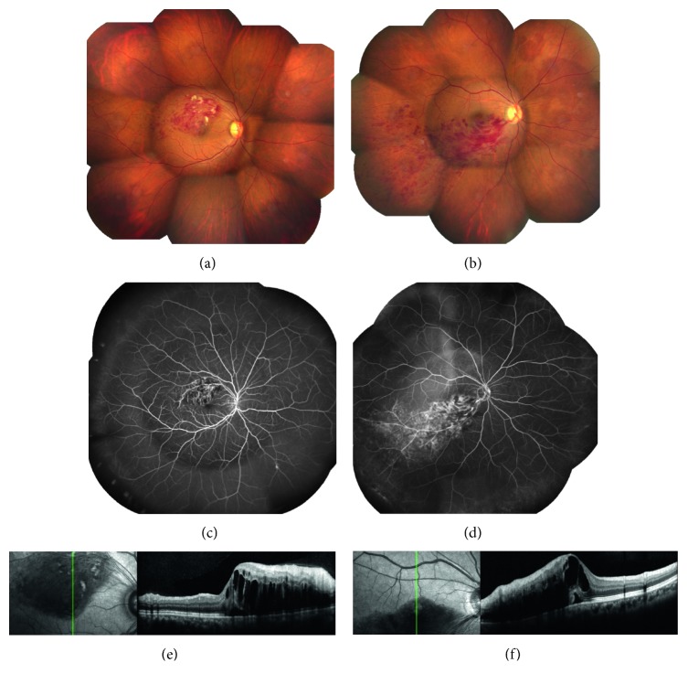
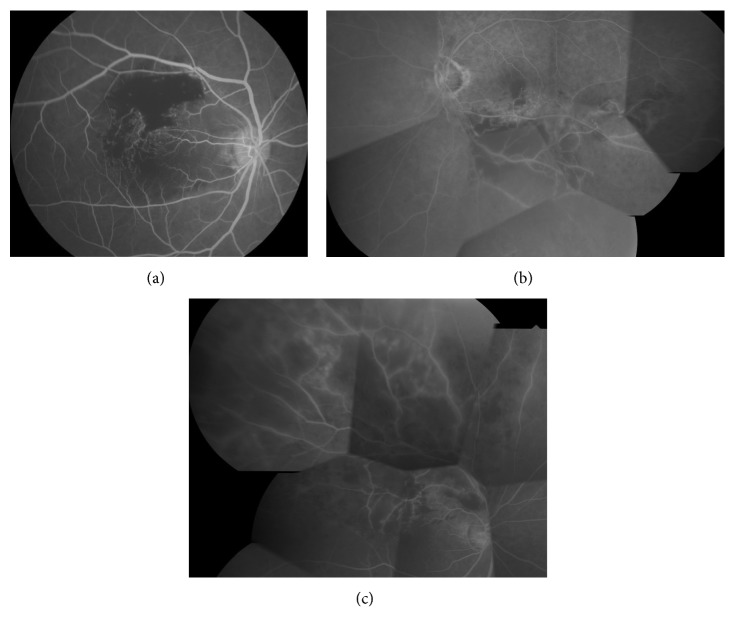
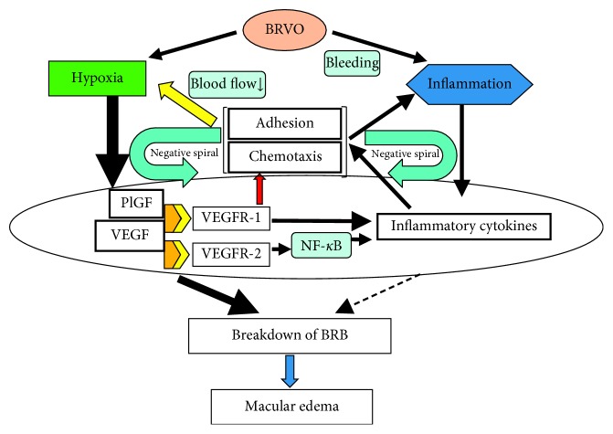
Branch retinal vein occlusion (BRVO) is a very common retinal vascular problem in patients with lifestyle-related diseases, such as hypertension and arteriosclerosis. In patients with BRVO, development of macular edema is the main cause of visual impairment. BRVO is still a controversial condition in many respects. Over the years, various methods such as laser photocoagulation have been tried to treat macular edema associated with BRVO, but the results were not satisfactory. After vascular endothelial growth factor (VEGF) was found to have an important role in the pathogenesis of macular edema in BRVO patients, treatment of this condition was revolutionized by development of anti-VEGF therapy. Although macular edema improves dramatically following intraocular injection of anti-VEGF agents, repeated recurrence and resistance of edema is a major problem in some BRVO patients. This suggests that factors or cytokines other than VEGF may be associated with inflammation and retinal hypoxia in BRVO and that the pathogenesis of macular edema is complicated. The present review assesses the role of various factors and cytokines in the pathogenesis of macular edema associated with BRVO. We present a mechanism that is not only plausible but should also be useful for developing new therapeutic strategies.


Figures

Figure 1
Figure 1
Color fundus photograph. (a) Macular branch retinal vein occlusion (BRVO). (b) Major BRVO. Fluorescein angiography. (c) Mild retinal hypoxia in macular BRVO. (d) Mild retinal hypoxia in major BRVO. Optical coherence tomography. (e) Macular edema and serous retinal detachment can occur in macular BRVO, even if retinal hypoxia is mild. (f) Macular edema and serous retinal detachment can also occur in major BRVO despite mild retinal hypoxia.
Figure 2
Figure 2
Vitreous fluid VEGF level and retinal nonperfusion in representative cases. Fluorescein angiograms. (a) Mild ischemia (15.6 pg/ml). The VEGF level is often 30–40 pg/ml in patients with mild nonperfusion. (b) Moderate ischemia (338 pg/ml). The VEGF level is often 300 pg/ml or higher when there is moderate nonperfusion. (c) Severe ischemia (2,570 pg/ml). The VEGF is often 2000–3000 pg/ml when nonperfusion is severe.
Figure 3
Figure 3
Pathogenesis of macular edema (hypothesis). BRVO not only causes retinal hypoxia but also produces inflammation secondary to retinal hemorrhage. As a result, expression of VEGF and inflammatory cytokines increases, resulting in disruption of the BRB with the development and progression of macular edema. Activation of VEGFR-1 by both VEGF and PlGF plays a role in recruitment of leukocytes and also upregulates expression of inflammatory cytokines. In addition, activation of VEGFR-2 by VEGF increases vascular permeability and enhances the expression of inflammatory cytokines such as MCP-1 and ICAM-1 via NF-κB, leading to chemotaxis and adhesion of leukocytes to the vascular endothelium along with a decrease of blood flow velocity. Reduction of blood flow velocity creates a positive feedback loop, which further exacerbates retinal hypoxia. Increased leukocyte chemotaxis and adhesion also enhances inflammation, creating another positive feedback loop. According to this hypothesis, the pathological mechanism becomes more complicated and the influence of inflammation increases as BRVO becomes chronic.

Similar articles

Cited by

References

    1. McIntosh R. L., Mohamed Q., Saw S. M., Wong T. Y. Interventions for branch retinal vein occlusion. Ophthalmology. 2007;114(5):835–854. doi: 10.1016/j.ophtha.2007.01.010. - DOI - PubMed
    1. Campochiaro P. A., Heier J. S., Feiner L., et al. Ranibizumab for macular edema following branch retinal vein occlusion. Ophthalmology. 2010;117(6):1102–1112. doi: 10.1016/j.ophtha.2010.02.021. - DOI - PubMed
    1. Christoffersen N. L. B., Larsen M. Pathophysiology and hemodynamics of branch retinal vein occlusion. Ophthalmology. 1999;106(11):2054–2062. doi: 10.1016/s0161-6420(99)90483-9. - DOI - PubMed
    1. Tsujikawa A., Sakamoto A., Ota M., et al. Serous retinal detachment associated with retinal vein occlusion. American Journal of Ophthalmology. 2010;149(2):291–301.e5. doi: 10.1016/j.ajo.2009.09.007. - DOI - PubMed
    1. Orth D. H., Patz A. Retinal branch vein occlusion. Survey of Ophthalmology. 1978;22(6):357–376. doi: 10.1016/0039-6257(78)90132-7. - DOI - PubMed

LinkOut - more resources