

Pigmented Basal Cell Carcinoma Masquerading as a Melanoma
- PMID: 31192074
- PMCID: PMC6551196
- DOI: 10.7759/cureus.4369
Pigmented Basal Cell Carcinoma Masquerading as a Melanoma
Abstract
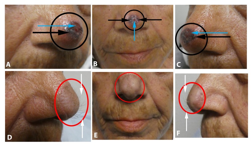
Basal cell carcinoma is the most common skin cancer. Pigmented basal cell carcinoma is an uncommon clinical presentation that can resemble a melanoma. We present the clinical and pathologic features of three individuals whose pigmented basal cell carcinomas masqueraded as melanomas. All of the patients were Hispanic and ranged in age from 63 years to 77 years. They presented with a pigmented lesion that was ultimately diagnosed as a pigmented basal cell carcinoma; one woman had a collision tumor consisting of a pigmented basal cell carcinoma and a seborrheic keratosis. All of the patients had their tumors removed using Mohs micrographic surgery, without recurrence. The clinical differential diagnosis of a black tumor-particularly in patients with darker skin types-should include pigmented basal cell carcinoma in addition to melanoma; a biopsy of the lesion will establish the diagnosis.
Keywords: basal; carcinoma; cell; collision; masquerading; melanoma; mimic; nodular; pigment; pigmented.
Conflict of interest statement
The authors have declared that no competing interests exist.
Figures


Similar articles
-
Morphologic Mimickers of Seborrheic Keratoses: Cutaneous Lesions Masquerading as Seborrheic Keratoses.Cureus. 2021 Oct 7;13(10):e18559. doi: 10.7759/cureus.18559. eCollection 2021 Oct. Cureus. 2021. PMID: 34765343 Free PMC article.
-
Eccrine porocarcinoma arising in a seborrheic keratosis evaluated with dermoscopy and treated with Mohs' technique.Int J Dermatol. 2003 Aug;42(8):653-7. doi: 10.1046/j.1365-4362.2003.01779.x. Int J Dermatol. 2003. PMID: 12890117
-
Pigmented basal cell cancer masquerading as superficial spreading malignant melanoma.Arch Dermatol. 1977 Jul;113(7):946-7. Arch Dermatol. 1977. PMID: 879817
-
Role of In Vivo Reflectance Confocal Microscopy in the Analysis of Melanocytic Lesions.Acta Dermatovenerol Croat. 2018 Apr;26(1):64-67. Acta Dermatovenerol Croat. 2018. PMID: 29782304 Review.
-
Spreading pigmented actinic keratosis: a review.J Am Acad Dermatol. 2010 Sep;63(3):499-506. doi: 10.1016/j.jaad.2009.07.026. Epub 2010 Mar 23. J Am Acad Dermatol. 2010. PMID: 20334953 Review.
Cited by
-
Clinicopathologic comparison of basal cell carcinoma among a diverse patient population in Los Angeles County.Skin Health Dis. 2024 Mar 24;4(4):e379. doi: 10.1002/ski2.379. eCollection 2024 Aug. Skin Health Dis. 2024. PMID: 39104648 Free PMC article.
-
Diagnostic dilemma in pigmented basal cell carcinoma: A case report.J Educ Health Promot. 2024 Jul 5;13:171. doi: 10.4103/jehp.jehp_83_24. eCollection 2024. J Educ Health Promot. 2024. PMID: 39268443 Free PMC article.
-
Dermoscopic features of neoplasms in skin of color: A review.Int J Womens Dermatol. 2021 Jan 19;7(2):145-151. doi: 10.1016/j.ijwd.2020.11.009. eCollection 2021 Mar. Int J Womens Dermatol. 2021. PMID: 33937480 Free PMC article. Review.
-
Clinical and Dermoscopic Patterns of Basal Cell Carcinoma and Its Mimickers in Skin of Color: A Practical Summary.Medicina (Kaunas). 2024 Aug 24;60(9):1386. doi: 10.3390/medicina60091386. Medicina (Kaunas). 2024. PMID: 39336428 Free PMC article. Review.
-
Basal cell carcinoma: Comprehensive clinical and histopathological aspects, novel imaging tools and therapeutic approaches (Review).Exp Ther Med. 2022 Jan;23(1):60. doi: 10.3892/etm.2021.10982. Epub 2021 Nov 18. Exp Ther Med. 2022. PMID: 34917186 Free PMC article. Review.
References
-
- Basal and squamous cell carcinoma. Garner KL, Rodney WM. Prim Care. 2000;27:447–458. - PubMed
-
- Annual report to the nation on the status of cancer, 1975-2001, with a special feature regarding survival. Jemal A, Clegg LX, Ward E, et al. Cancer. 2004;101:3–27. - PubMed
-
- The epidemiology of skin cancer. Diepgen TL, Mahler V. Br J Dermatol. 2002;146:1–6. - PubMed
-
- The pigmented basal cell epithelioma. Zelickson AS. Arch Derm. 1967;96:524–527. - PubMed
-
- A histologic and electron microscopic study of a pigmenting basal cell epithelioma. Zelickson AS, Goltz RW, Hartmann JF. J Invest Dermatol. 1961;36:299–302. - PubMed
Publication types
LinkOut - more resources
Full Text Sources