

An in vivo Like Micro-Carcinoma Model
- PMID: 31192122
- PMCID: PMC6540606
- DOI: 10.3389/fonc.2019.00410
An in vivo Like Micro-Carcinoma Model
Abstract
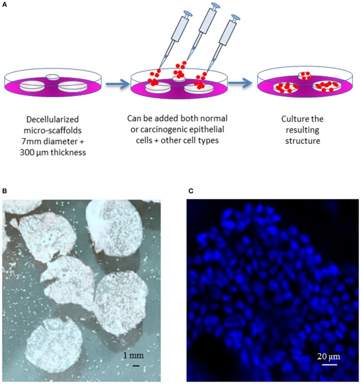
We here present a novel micro-system which allows to reconstitute an in vivo lung carcinoma where the various constituting epithelial and/or stromal structural and/or cellular components can be incorporated at will. In contrast to various "organs on a chip" the model is based on the observation that in nature, epithelial cells are always supported by a connective tissue or stroma. The model is based on acellular micro-scaffolds of microscopic dimensions which enable seeded cells to obtain gases and nutrients through diffusion thus avoiding the need for vascularization. As a proof of concept, we show that in this model, Calu-3 cells can form a well-organized, continuous, polarized, one-layer epithelium lining the stromal derived alveolar cavities, and express a different pattern of tumor-related genes than when grown as standard monolayer cultures on plastic culture dishes. To our knowledge, this model, introduces for the first time a system where the function of carcinogenic cells can be tested in vitro in an environment that closely mimics the natural in vivo situation.
Keywords: carcinoma; decellularized scaffolds; tumor micro-culture; tumor microenvironment; tumor model.
Figures
Similar articles
-
Paracrine Signaling from a Three-Dimensional Model of Bladder Carcinoma and from Normal Bladder Switch the Phenotype of Stromal Fibroblasts.Cancers (Basel). 2021 Jun 14;13(12):2972. doi: 10.3390/cancers13122972. Cancers (Basel). 2021. PMID: 34198488 Free PMC article.
-
Organ-specific scaffolds for in vitro expansion, differentiation, and organization of primary lung cells.Tissue Eng Part C Methods. 2011 Aug;17(8):861-70. doi: 10.1089/ten.tec.2010.0717. Epub 2011 May 19. Tissue Eng Part C Methods. 2011. PMID: 21595544
-
Beta Cells Secrete Significant and Regulated Levels of Insulin for Long Periods when Seeded onto Acellular Micro-Scaffolds.Tissue Eng Part A. 2015 Nov;21(21-22):2691-702. doi: 10.1089/ten.TEA.2014.0711. Tissue Eng Part A. 2015. PMID: 26416226
-
Tumor-stroma interactions directing phenotype and progression of epithelial skin tumor cells.Differentiation. 2002 Dec;70(9-10):486-97. doi: 10.1046/j.1432-0436.2002.700903.x. Differentiation. 2002. PMID: 12492491 Review.
-
Organ reconstruction: Dream or reality for the future.Biomed Mater Eng. 2017;28(s1):S121-S127. doi: 10.3233/BME-171633. Biomed Mater Eng. 2017. PMID: 28372287 Review.
Cited by
-
Paracrine Signaling from a Three-Dimensional Model of Bladder Carcinoma and from Normal Bladder Switch the Phenotype of Stromal Fibroblasts.Cancers (Basel). 2021 Jun 14;13(12):2972. doi: 10.3390/cancers13122972. Cancers (Basel). 2021. PMID: 34198488 Free PMC article.
-
Assessment of Antitumor and Antiproliferative Efficacy and Detection of Protein-Protein Interactions in Cancer Cells from 3D Tumor Spheroids.Curr Protoc. 2022 Oct;2(10):e569. doi: 10.1002/cpz1.569. Curr Protoc. 2022. PMID: 36286844 Free PMC article.
References
LinkOut - more resources
Full Text Sources
Research Materials