

Rational Combination of Parvovirus H1 With CTLA-4 and PD-1 Checkpoint Inhibitors Dampens the Tumor Induced Immune Silencing
- PMID: 31192129
- PMCID: PMC6546938
- DOI: 10.3389/fonc.2019.00425
Rational Combination of Parvovirus H1 With CTLA-4 and PD-1 Checkpoint Inhibitors Dampens the Tumor Induced Immune Silencing
Abstract
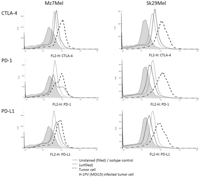
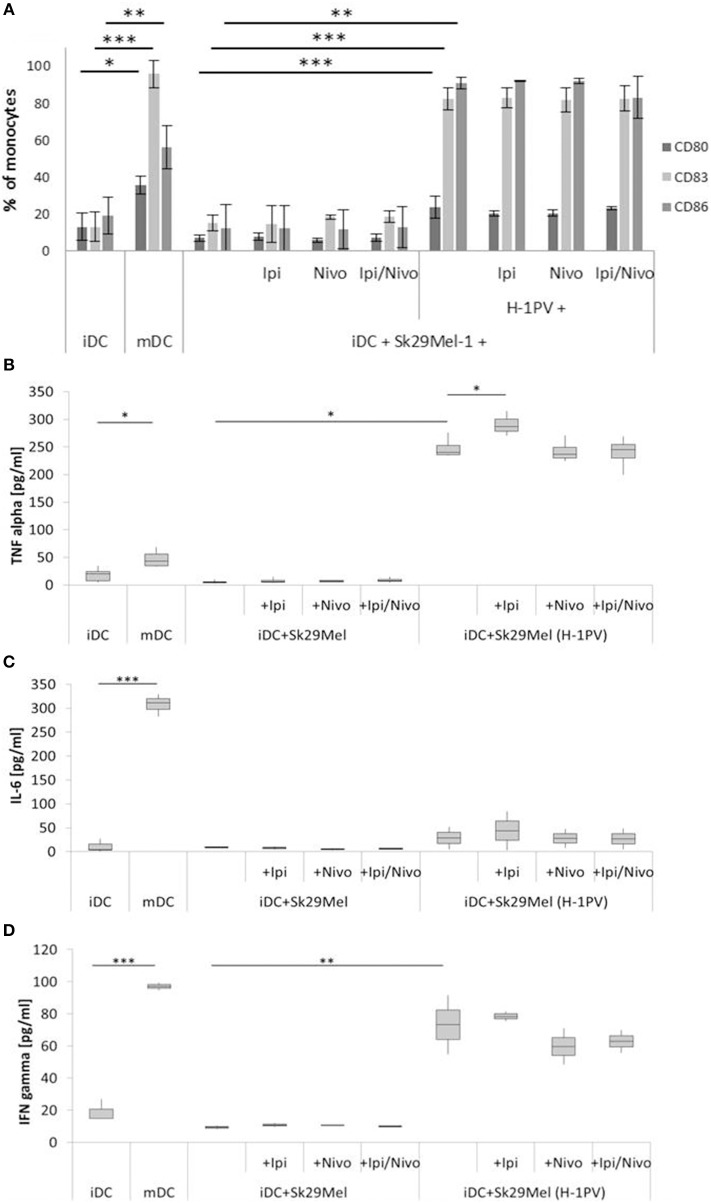
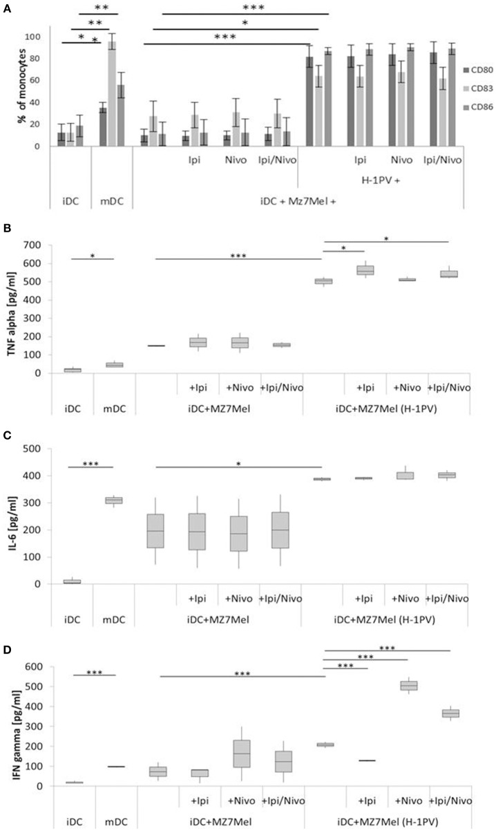
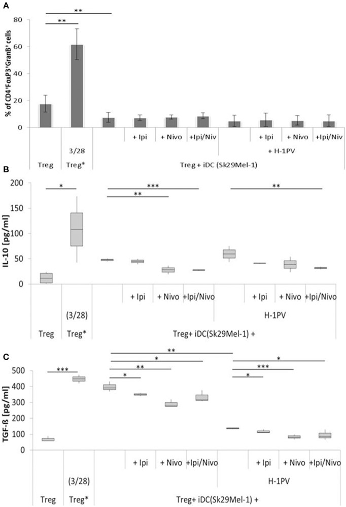
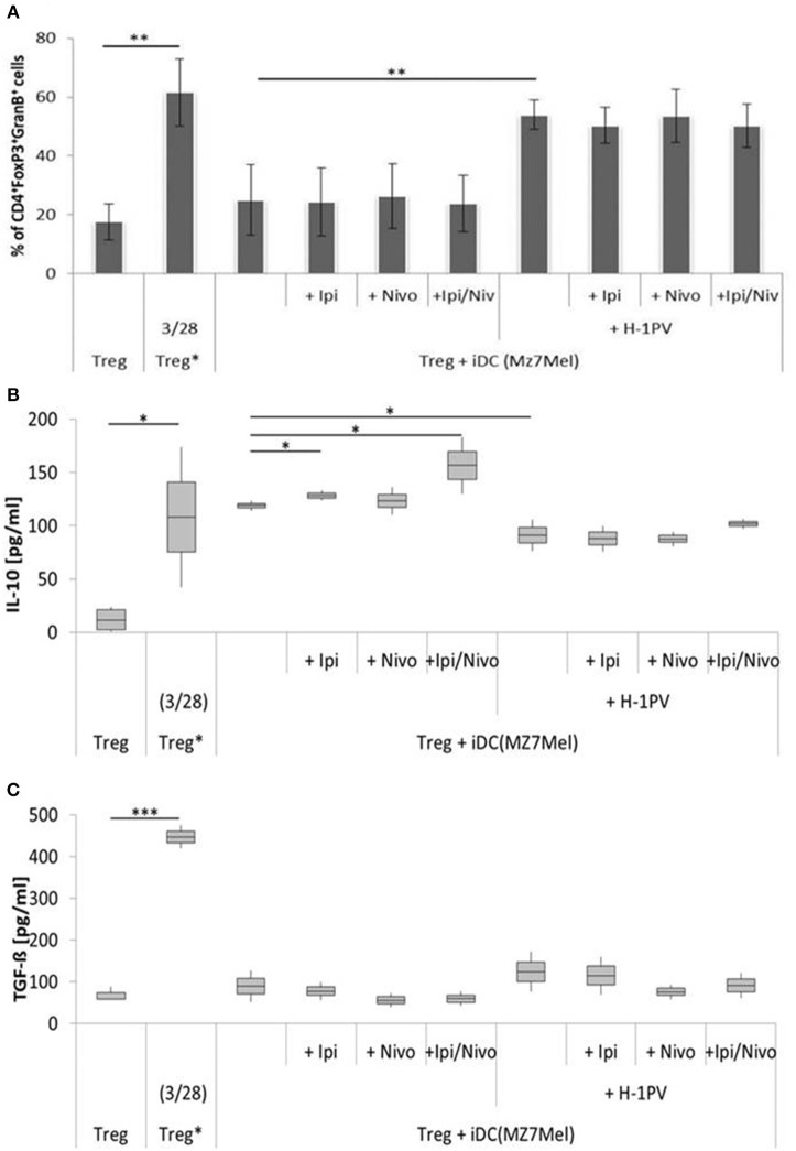
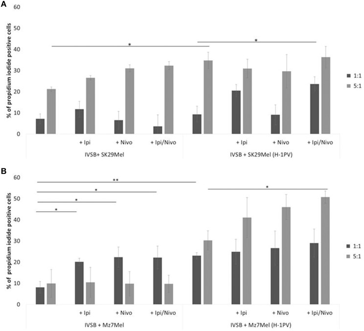
The recent therapeutic success of immune checkpoint inhibitors in the treatment of advanced melanoma highlights the potential of cancer immunotherapy. Oncolytic virus-based therapies may further improve the outcome of these cancer patients. A human ex vivo melanoma model was used to investigate the oncolytic parvovirus H-1 (H-1PV) in combination with ipilimumab and/or nivolumab. The effect of this combination on activation of human T lymphocytes was demonstrated. Expression of CTLA-4, PD-1, and PD-L1 immune checkpoint proteins was upregulated in H-1PV-infected melanoma cells. Nevertheless, maturation of antigen presenting cells such as dendritic cells was triggered by H-1PV infected melanoma cells. Combining H-1PV with checkpoint inhibitors, ipilimumab enhanced TNFα release during maturation of dendritic cells; nivolumab increased the amount of IFNγ release. H-1PV mediated reduction of regulatory T cell activity was demonstrated by lower TGF-ß levels. The combination of ipilimumab and nivolumab resulted in a further decline of TGF-ß levels. Similar results were obtained regarding the activation of cytotoxic T cells. H-1PV infection alone and in combination with both checkpoint inhibitors caused strong activation of CTLs, which was reflected by an increased number of CD8+GranB+ cells and increased release of granzyme B, IFNγ, and TNFα. Our data support the concept of a treatment benefit from combining oncolytic H-1PV with the checkpoint inhibitors ipilimumab and nivolumab, with nivolumab inducing stronger effects on cytotoxic T cells, and ipilimumab strengthening T lymphocyte activity.
Keywords: H-1PV; immune cells; immunotherapy; ipilimumab; melanoma; nivolumab.
Figures
Similar articles
-
The Next Immune-Checkpoint Inhibitors: PD-1/PD-L1 Blockade in Melanoma.Clin Ther. 2015 Apr 1;37(4):764-82. doi: 10.1016/j.clinthera.2015.02.018. Epub 2015 Mar 29. Clin Ther. 2015. PMID: 25823918 Free PMC article. Review.
-
Activation of the human immune system by chemotherapeutic or targeted agents combined with the oncolytic parvovirus H-1.BMC Cancer. 2011 Oct 26;11:464. doi: 10.1186/1471-2407-11-464. BMC Cancer. 2011. PMID: 22029859 Free PMC article.
-
Influence of the oncolytic parvovirus H-1, CTLA-4 antibody tremelimumab and cytostatic drugs on the human immune system in a human in vitro model of colorectal cancer cells.Onco Targets Ther. 2013 Aug 20;6:1119-27. doi: 10.2147/OTT.S49371. eCollection 2013. Onco Targets Ther. 2013. PMID: 23986643 Free PMC article.
-
Activation of the human immune system via toll-like receptors by the oncolytic parvovirus H-1.Int J Cancer. 2013 Jun 1;132(11):2548-56. doi: 10.1002/ijc.27938. Epub 2012 Nov 29. Int J Cancer. 2013. PMID: 23151948
-
Combination therapy with PD-1 or PD-L1 inhibitors for cancer.Int J Clin Oncol. 2020 May;25(5):818-830. doi: 10.1007/s10147-019-01548-1. Epub 2019 Sep 23. Int J Clin Oncol. 2020. PMID: 31549270 Review.
Cited by
-
Virotherapy in Germany-Recent Activities in Virus Engineering, Preclinical Development, and Clinical Studies.Viruses. 2021 Jul 21;13(8):1420. doi: 10.3390/v13081420. Viruses. 2021. PMID: 34452286 Free PMC article. Review.
-
Virotherapy combined with anti-PD-1 transiently reshapes the tumor immune environment and induces anti-tumor immunity in a preclinical PDAC model.Front Immunol. 2023 Jan 16;13:1096162. doi: 10.3389/fimmu.2022.1096162. eCollection 2022. Front Immunol. 2023. PMID: 36726983 Free PMC article.
-
Research Status and Outlook of PD-1/PD-L1 Inhibitors for Cancer Therapy.Drug Des Devel Ther. 2020 Sep 8;14:3625-3649. doi: 10.2147/DDDT.S267433. eCollection 2020. Drug Des Devel Ther. 2020. PMID: 32982171 Free PMC article. Review.
-
Oncolytic virotherapy reverses the immunosuppressive tumor microenvironment and its potential in combination with immunotherapy.Cancer Cell Int. 2021 May 13;21(1):262. doi: 10.1186/s12935-021-01972-2. Cancer Cell Int. 2021. PMID: 33985527 Free PMC article. Review.
-
Oncolytic measles vaccines encoding PD-1 and PD-L1 checkpoint blocking antibodies to increase tumor-specific T cell memory.Mol Ther Oncolytics. 2021 Nov 29;24:43-58. doi: 10.1016/j.omto.2021.11.020. eCollection 2022 Mar 17. Mol Ther Oncolytics. 2021. PMID: 34977341 Free PMC article.
References
-
- Menshawy A, Eltonob AA, Barkat SA, Ghanem A, Mniesy MM, Mohamed I, et al. . Nivolumab monotherapy or in combination with ipilimumab for metastatic melanoma: systematic review and meta-analysis of randomized-controlled trials. Melanoma Res. (2018) 28:371–9. 10.1097/cmr.0000000000000467 - DOI - PubMed
-
- Moehler M, Blechacz B, Weiskopf N, Zeidler M, Stremmel W, Rommelaere J, et al. Effective infection, apoptotic cell killing and gene transfer of human hepatoma cells but not primary hepatocytes by parvovirus H1 and derived vectors. Cancer Gene Ther. (2001) 8:158–67. 10.1038/sj.cgt.7700288 - DOI - PubMed
LinkOut - more resources
Full Text Sources
Other Literature Sources
Research Materials