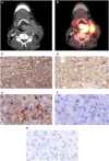
CT Texture Analysis-Correlations With Histopathology Parameters in Head and Neck Squamous Cell Carcinomas
- PMID: 31192138
- PMCID: PMC6546809
- DOI: 10.3389/fonc.2019.00444
CT Texture Analysis-Correlations With Histopathology Parameters in Head and Neck Squamous Cell Carcinomas
Abstract
Introduction: Texture analysis is an emergent imaging technique to quantify heterogeneity in radiological images. It is still unclear whether this technique is capable to reflect tumor microstructure. The present study sought to correlate histopathology parameters with texture features derived from contrast-enhanced CT images in head and neck squamous cell carcinomas (HNSCC). Materials and Methods: Twenty-eight patients with histopathological proven HNSCC were retrospectively analyzed. In every case EGFR, VEGF, Hif1-alpha, Ki67, p53 expression derived from immunhistochemical specimen were semiautomatically calculated. Furthermore, mean cell count was estimated. Texture analysis was performed on contrast-enhanced CT images as a whole lesion measurement. Spearman's correlation analysis was performed, adjusted with Benjamini-Hochberg correction for multiple tests. Results: Several texture features correlated with histopathological parameters. After correction only CT texture joint entropy and CT entropy correlation with Hif1-alpha expression remained statistically significant (ρ = -0.60 and ρ = -0.50, respectively). Conclusions: CT texture joint entropy and CT entropy were associated with Hif1-alpha expression in HNSCC and might be able to reflect hypoxic areas in this entity.
Keywords: CT; HNSCC; Hif1-alpha; Ki67; texture analysis.
Figures


Similar articles
-
ADC-histogram analysis in head and neck squamous cell carcinoma. Associations with different histopathological features including expression of EGFR, VEGF, HIF-1α, Her 2 and p53. A preliminary study.Magn Reson Imaging. 2018 Dec;54:214-217. doi: 10.1016/j.mri.2018.07.013. Epub 2018 Sep 4. Magn Reson Imaging. 2018. PMID: 30189236
-
Associations between Histogram Analysis Parameters Derived from DCE-MRI and Histopathological Features including Expression of EGFR, p16, VEGF, Hif1-alpha, and p53 in HNSCC.Contrast Media Mol Imaging. 2019 Jan 2;2019:5081909. doi: 10.1155/2019/5081909. eCollection 2019. Contrast Media Mol Imaging. 2019. PMID: 30718984 Free PMC article.
-
Histogram Analysis Parameters Derived from Conventional T1- and T2-Weighted Images Can Predict Different Histopathological Features Including Expression of Ki67, EGFR, VEGF, HIF-1α, and p53 and Cell Count in Head and Neck Squamous Cell Carcinoma.Mol Imaging Biol. 2019 Aug;21(4):740-746. doi: 10.1007/s11307-018-1283-y. Mol Imaging Biol. 2019. PMID: 30284155
-
Associations Between [18F]FDG-PET and Complex Histopathological Parameters Including Tumor Cell Count and Expression of KI 67, EGFR, VEGF, HIF-1α, and p53 in Head and Neck Squamous Cell Carcinoma.Mol Imaging Biol. 2019 Apr;21(2):368-374. doi: 10.1007/s11307-018-1223-x. Mol Imaging Biol. 2019. PMID: 29931433
-
Relationships between histogram analysis of ADC values and complex 18F-FDG-PET parameters in head and neck squamous cell carcinoma.PLoS One. 2018 Sep 6;13(9):e0202897. doi: 10.1371/journal.pone.0202897. eCollection 2018. PLoS One. 2018. PMID: 30188926 Free PMC article.
Cited by
-
CT texture analysis of tonsil cancer: Discrimination from normal palatine tonsils.PLoS One. 2021 Aug 11;16(8):e0255835. doi: 10.1371/journal.pone.0255835. eCollection 2021. PLoS One. 2021. PMID: 34379652 Free PMC article.
-
CT texture analysis and node-RADS CT score of mediastinal lymph nodes - diagnostic performance in lung cancer patients.Cancer Imaging. 2022 Dec 26;22(1):75. doi: 10.1186/s40644-022-00506-x. Cancer Imaging. 2022. PMID: 36567339 Free PMC article.
-
Associations between dynamic-contrast enhanced MRI with histopathological features in atypical HCC using spatial co-registration with biopsy.Am J Transl Res. 2025 Apr 15;17(4):2967-2975. doi: 10.62347/PJYE7877. eCollection 2025. Am J Transl Res. 2025. PMID: 40384997 Free PMC article.
-
Computed Tomography Embolus Texture Analysis as a Prognostic Marker of Acute Pulmonary Embolism.Angiology. 2023 May;74(5):461-471. doi: 10.1177/00033197221111862. Epub 2022 Aug 16. Angiology. 2023. PMID: 35973807 Free PMC article.
-
Impact of CT texture analysis on complication rate in CT-guided liver biopsies.Clin Exp Hepatol. 2024 Mar;10(1):72-78. doi: 10.5114/ceh.2024.134141. Epub 2024 Jan 4. Clin Exp Hepatol. 2024. PMID: 38765907 Free PMC article.
References
-
- Surov A, Stumpp P, Meyer HJ, Gawlitza M, Höhn AK, Boehm A, et al. . Simultaneous (18)F-FDG-PET/MRI: associations between diffusion, glucose metabolism and histopathological parameters in patients with head and neck squamous cell carcinoma. Oral Oncol. (2016) 58:14–20. 10.1016/j.oraloncology.2016.04.009 - DOI - PubMed
-
- Meyer HJ, Schob S, Münch B, Frydrychowicz C, Garnov N, Quäschling U, et al. . Histogram analysis of T1-weighted, T2-weighted, and postcontrast T1-weighted images in primary CNS lymphoma: correlations with histopathological findings-a preliminary study. Mol Imaging Biol. (2018) 20:318–23. 10.1007/s11307-017-1115-5 - DOI - PubMed
LinkOut - more resources
Full Text Sources
Research Materials
Miscellaneous

