. 2020 Apr;66(4):423-432.
doi: 10.1097/MAT.0000000000001018.

Low-Resistance, Concentric-Gated Pediatric Artificial Lung for End-Stage Lung Failure

Affiliations

Low-Resistance, Concentric-Gated Pediatric Artificial Lung for End-Stage Lung Failure

Alex J Thompson et al. ASAIO J. 2020 Apr.

Abstract

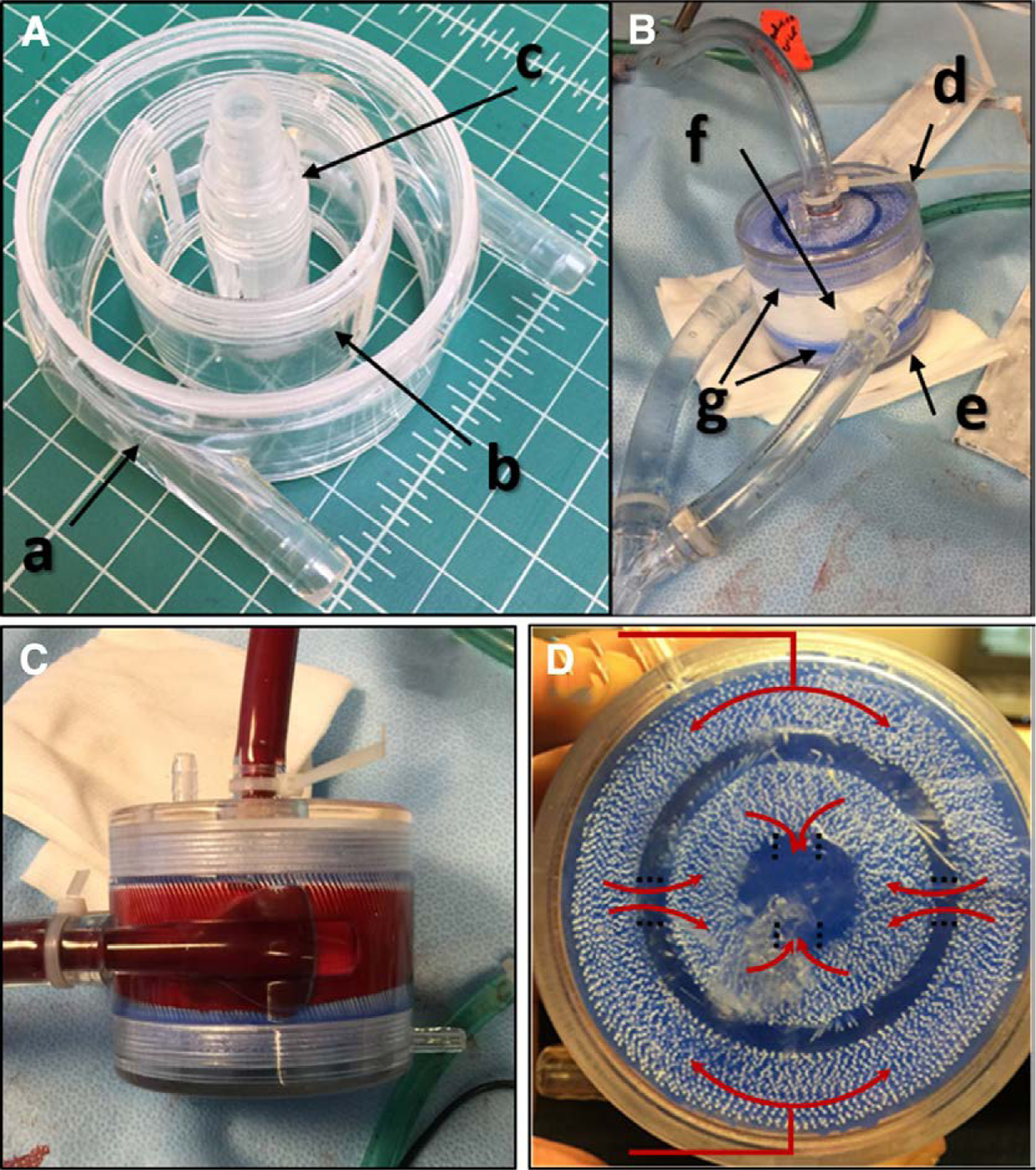
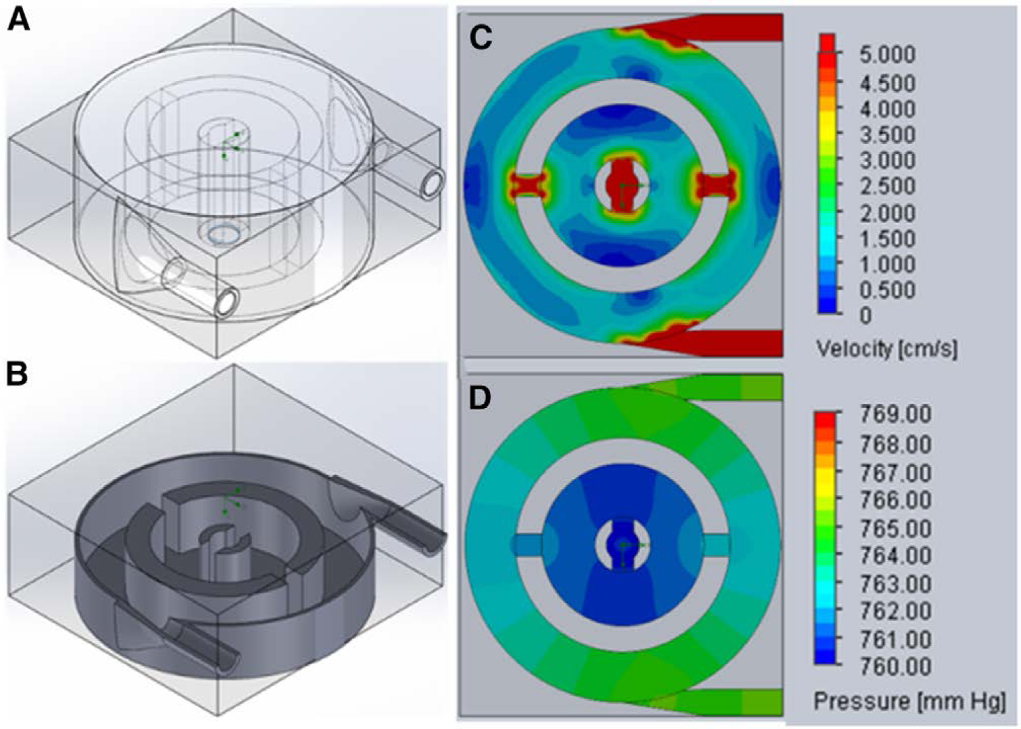
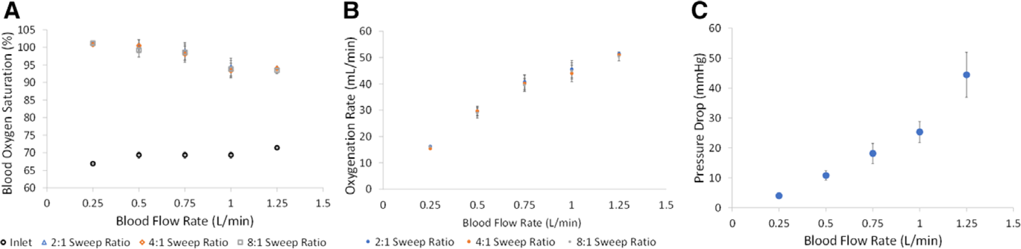
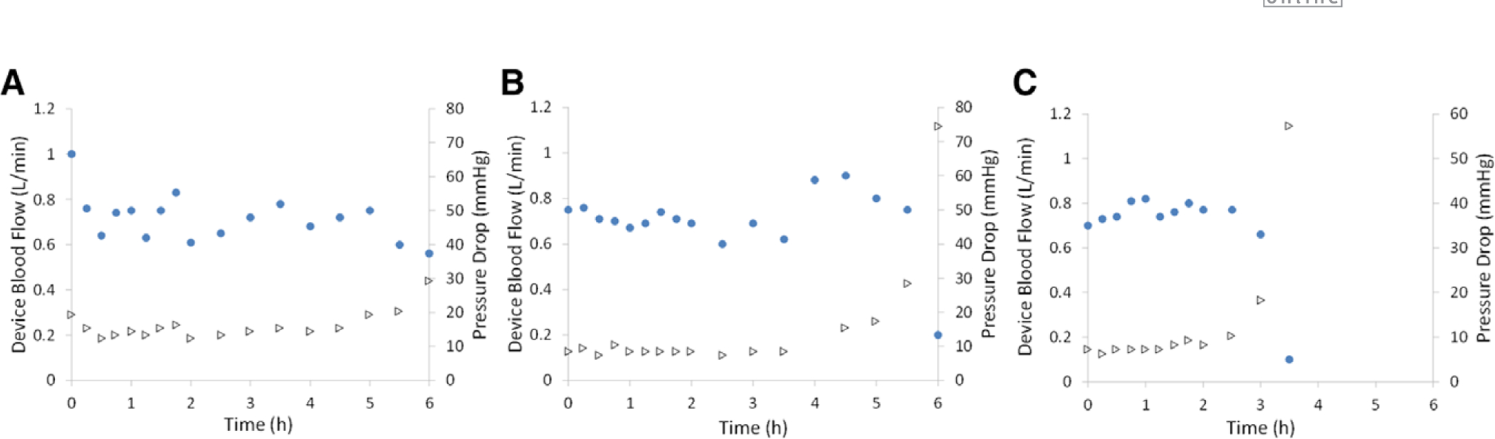
Children with end-stage lung failure awaiting lung transplant would benefit from improvements in artificial lung technology allowing for wearable pulmonary support as a bridge-to-transplant therapy. In this work, we designed, fabricated, and tested the Pediatric MLung-a dual-inlet hollow fiber artificial lung based on concentric gating, which has a rated flow of 1 L/min, and a pressure drop of 25 mm Hg at rated flow. This device and future iterations of the current design are designed to relieve pulmonary arterial hypertension, provide pulmonary support, reduce ventilator-associated injury, and allow for more effective therapy of patients with end-stage lung disease, including bridge-to-transplant treatment.


Conflict of interest statement

Disclosure: The authors have no conflicts of interest to report.

Figures

Figure 1.
Figure 1.
Basic device topology of the Pediatric MLung. A, B: The components the comprise the device include (a) outer housing, (b) gate insert, (c) blood outlet, (d, e) end caps, (f) fiber bundles, and (g) the blue silicone potting. C: Side view of a Pediatric MLung; the fibers are oriented vertically within the housing in this image. D: Top view of a Pediatric MLung with illustration of the blood flow paths (red arrows).
Figure 2.
Figure 2.
Isometric views of the 3D model (A) and a cross-section through a midplane (B) of the pediatric MLung having 3 cm fiber length. Velocity (C) and pressure (D) profiles through the mid-plane of the Pediatric MLung were generated with Solidworks Flow Simulation. 3D, three-dimensional.
Figure 3.
Figure 3.
Oxygenation (A, B) and pressure drop (C) of pediatric MLung during benchtop blood flow testing (n = 6).
Figure 4.
Figure 4.
Device flow rate (circles) and corresponding pressure drop (triangles) during 6 hours of arteriovenous blood flow from adult sheep. Three Pediatric MLungs were tested (AC) for 6 hours or until device flow was reduced to 5% of initial flow.
Figure 5.
Figure 5.
Systemic ACT (circles) and device resistance (triangles) during 6 hours of arteriovenous blood flow from adult sheep. Three Pediatric MLungs were tested (AC) for 6 hours or until device flow was reduced to 5% of initial flow. ACT, activated clotting time.
Figure 6.
Figure 6.
Platelet count (circles) and WBC count (triangles) during 6 hours of arteriovenous blood flow from adult sheep. Three Pediatric MLungs were tested (AC) for 6 hours or until device flow was reduced to 5% of initial flow. WBC, white blood cell.
Figure 7.
Figure 7.
CO2 removal efficiency (circles) at varied sweep flow (triangles) of three Pediatric MLungs that were tested for 6 hours or until device flow was reduced to 5% of initial flow. Datapoints not gathered were due to dilution of the sample by sweep gas making the collected CO2 concentration below the detection limit of the blood gas machine (average ± SEM, n = 3, *n = 2, no error bar: n = 1). CO2, carbon dioxide; SEM, standard error mean.
Figure 8.
Figure 8.
Device flow rate (diamonds) and corresponding pressure drop (squares) during 6 hours of blood flow from adult sheep. Three Pediatric MLungs were tested (AC) for 6 hours (with exception of plot C, in which the experiment was ended voluntarily after occlusion of the rPA did not result in blood flow of at least 0.75 L/min). Oxygenation rate was measured throughout each experiment (D). rPA, right pulmonary artery.

References

    1. Valapour M, Skeans MA, Smith JM, et al.: OPTN/SRTR 2015 annual data report: lung. Am J Transplant 17(suppl 1): 357–424, 2017. - PubMed
    1. Alghanem F, Davis RP, Bryner BS, et al.: The implantable pediatric artificial lung: interim report on the development of an end-stage lung failure model. ASAIO J 61: 453–458, 2015. - PMC - PubMed
    1. Boston US, Fehr J, Gazit AZ, Eghtesady P: Paracorporeal lung assist device: an innovative surgical strategy for bridging to lung transplant in an infant with severe pulmonary hypertension caused by alveolar capillary dysplasia. J Thorac Cardiovasc Surg 146: e42–e43, 2013. - PubMed
    1. Hoganson DM, Gazit AZ, Boston US, et al.: Paracorporeal lung assist devices as a bridge to recovery or lung transplantation in neonates and young children. J Thorac Cardiovasc Surg 147: 420–426, 2014. - PubMed
    1. Alghanem F, Bryner BS, Jahangir EM, et al.: Pediatric artificial lung: a low-resistance pumpless artificial lung alleviates an acute lamb model of increased right ventricle afterload. ASAIO J 63: 223–228, 2017. - PMC - PubMed

Publication types