. 2019 Jul;40(6):736-744.
doi: 10.1097/MAO.0000000000002164.

Intracochlear Pressure Transients During Cochlear Implant Electrode Insertion: Effect of Micro-mechanical Control on Limiting Pressure Trauma

Affiliations

Intracochlear Pressure Transients During Cochlear Implant Electrode Insertion: Effect of Micro-mechanical Control on Limiting Pressure Trauma

Renee M Banakis Hartl et al. Otol Neurotol. 2019 Jul.

Abstract

Hypothesis: Use of micro-mechanical control during cochlear implant (CI) electrode insertion will result in reduced number and magnitude of pressure transients when compared with standard insertion by hand.

Introduction: With increasing focus on hearing preservation during CI surgery, atraumatic electrode insertion is of the utmost importance. It has been established that large intracochlear pressure spikes can be generated during the insertion of implant electrodes. Here, we examine the effect of using a micro-mechanical insertion control tool on pressure trauma exposures during implantation.

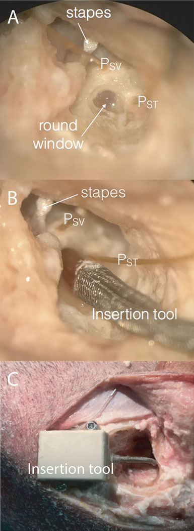
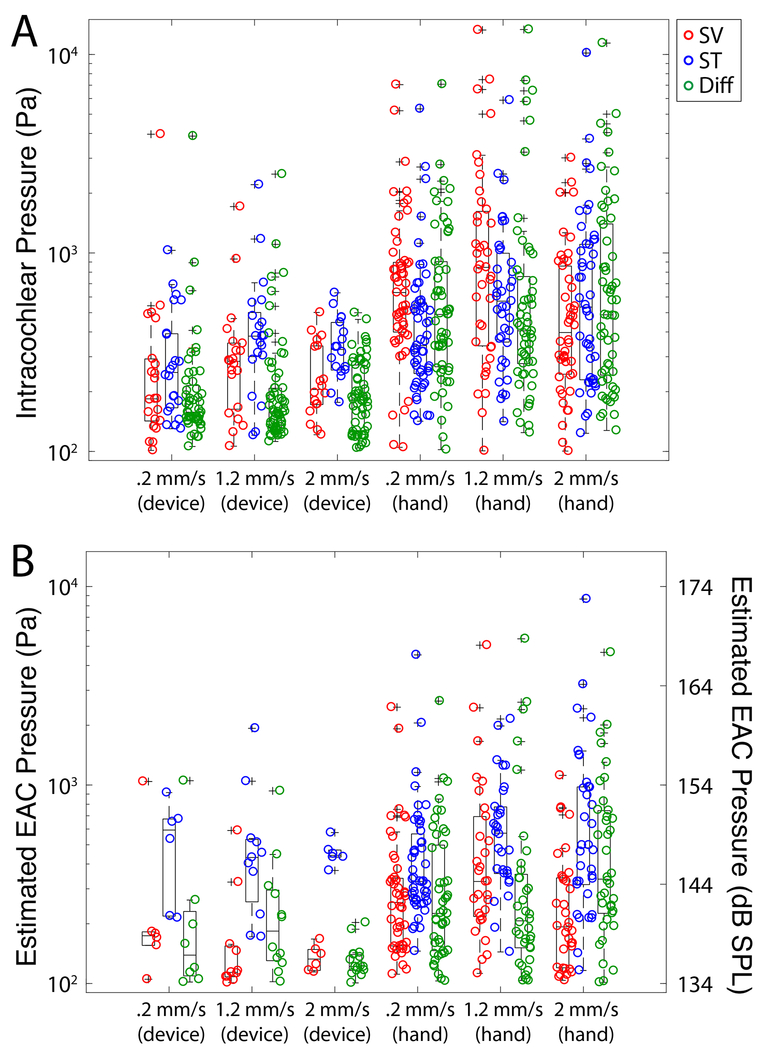
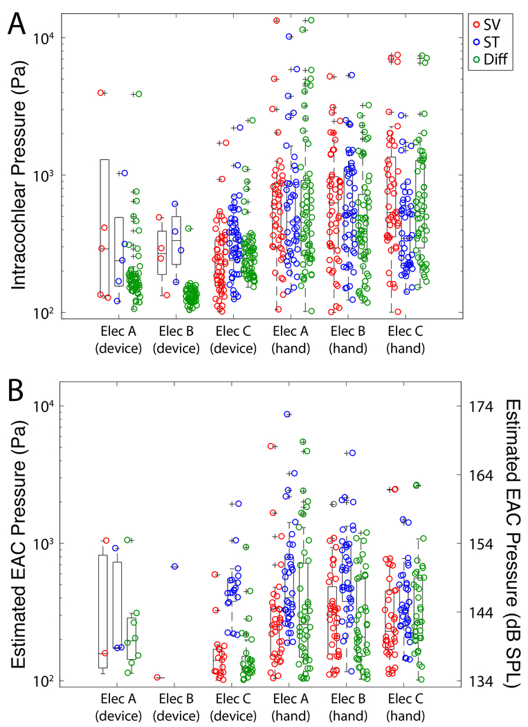
Methods: Human cadaveric heads were surgically prepared with an extended facial recess. Electrodes from three manufacturers were placed both by using a micro-mechanical control tool and by hand. Insertions were performed at three different rates: 0.2 mm/s, 1.2 mm/s, and 2 mm/s (n = 20 each). Fiber-optic sensors measured pressures in scala vestibuli and tympani.

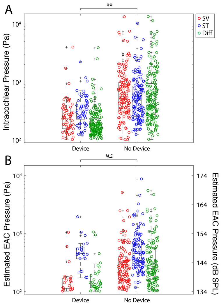
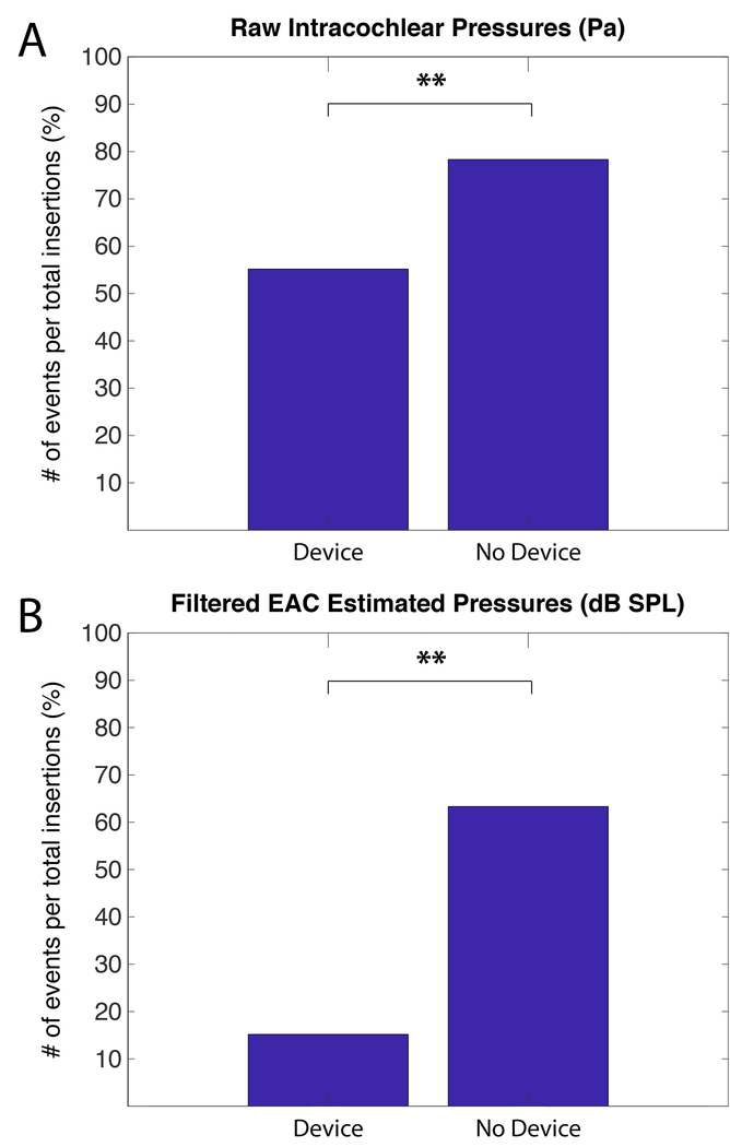
Results: Electrode insertion produced pressure transients up to 174 dB SPL. ANOVA revealed that pressures were significantly lower when using the micro-mechanical control device compared with insertion by hand (p << 0.001). No difference was noted across electrode type or speed. Chi-square analysis showed a significantly lower proportion of insertions contained pressure spikes when the control system was used (p << 0.001).

Conclusion: Results confirm previous data that suggest CI electrode insertion can cause pressure transients with intensities similar to those elicited by high-level sounds. Results suggest that the use of a micro-mechanical insertion control system may mitigate trauma from pressure events, both by reducing the amplitude and the number of pressure spikes resulting from CI electrode insertion.


Conflict of interest statement

Conflict of Interest Statement: CK and MRH are co-founders of iotaMotion, Inc.

Figures

Figure 1:
Figure 1:
Photomicrograph of the right ear in a single specimen during data collection. Through the extended facial recess, the anatomical landmarks (stapes and round window niche) can be visualized. Pressure probes (PSV and PST) were placed in cochleostomies in scala vestibuli and tympani (A). The insertion tool guide sheath was placed into the round window niche to direct implant electrode placement (B). The insertion tool unit secured by bone screws at edge of mastoid cavity with electrode coupled and housed within enclosure and guide sheath (C).
Figure 2:
Figure 2:
Mean baseline stapes velocity (VStap), scala vestibuli pressure (PSV), scala tympani pressure (PST), and differential pressure (PDiff; PSV-PST) transfer function magnitudes to air-conducted stimuli. Responses recorded in specimens are shown normalized to the SPL recorded in the ear canal (PEAC) and are superimposed onto the 95% CI and range of responses (gray bands) observed previously,. Colored bands indicate +/− standard error of the mean.
Figure 3:
Figure 3:
Summary of peak sound pressure levels observed in all specimens during all electrode insertions. Unfiltered peak intracochlear pressure measurements (A) and estimated EAC pressures (B) are shown for each pressure recording as a function of recording location. Box plots represent the median +/−25% of the range of pressures observed, whiskers show the full range of the estimated distribution, and +’s mark outliers. Significant differences between groups are indicated with asterisks (* p<0.05, ** p<0.01).
Figure 4:
Figure 4:
Summary of peak sound pressure levels by insertion rate. Unfiltered peak intracochlear pressure measurements (A) and estimated EAC pressures (B) are shown for each pressure recording as a function of insertion speed. Box plots represent the median +/−25% of the range of pressures observed, whiskers show the full range of the estimated distribution, and +’s mark outliers. No significant differences between groups were noted.
Figure 5:
Figure 5:
Summary of peak sound pressure levels by electrode type. Unfiltered peak intracochlear pressure measurements (A) and estimated EAC pressures (B) are shown for each pressure recording as a function of electrode type. Box plots represent the median +/−25% of the range of pressures observed, whiskers show the full range of the estimated distribution, and +’s mark outliers. No significant differences between groups were noted.
Figure 6:
Figure 6:
Percent of insertions with a significant pressure event by insertion duration. Chi-squared analysis with Bonferroni correction revealed a significant pattern of fewer exposures with the use of the micromechanical control device. Significant differences between groups are indicated with asterisks (* p<0.05, ** p<0.01).

Similar articles

Cited by

References

    1. Roland JT, Gantz BJ, Waltzman SB, Parkinson AJ, Multicenter Clinical Trial Group. United States multicenter clinical trial of the cochlear nucleus hybrid implant system. Laryngoscope. 2016;126(1):175–181. - PMC - PubMed
    1. Gifford RH, Grantham DW, Sheffield SW, Davis TJ, Dwyer R, Dorman MF. Localization and interaural time difference (ITD) thresholds for cochlear implant recipients with preserved acoustic hearing in the implanted ear. Hear Res. 2014;312:28–37. - PMC - PubMed
    1. Brown RF, Hullar TE, Cadieux JH, Chole RA. Residual hearing preservation after pediatric cochlear implantation. Otol Neurotol. 2010;31(8):1221–1226. - PMC - PubMed
    1. Dorman MF, Gifford RH, Spahr AJ, McKarns SA. The benefits of combining acoustic and electric stimulation for the recognition of speech, voice and melodies. Audiol Neurootol. 2008;13(2):105–112. - PMC - PubMed
    1. Turner CW, Gantz BJ, Vidal C, Behrens A, Henry BA. Speech recognition in noise for cochlear implant listeners: benefits of residual acoustic hearing. J Acoust Soc Am. 2004;115(4):1729–1735. - PubMed

Publication types