. 2019 Jun;98(24):e15910.
doi: 10.1097/MD.0000000000015910.

Incidence of recurrent fungal keratitis after primary keratoplasty and visual outcome and prognosis after intervention for the recurrence

Affiliations

Incidence of recurrent fungal keratitis after primary keratoplasty and visual outcome and prognosis after intervention for the recurrence

Yuerong Gong et al. Medicine (Baltimore). 2019 Jun.

Abstract

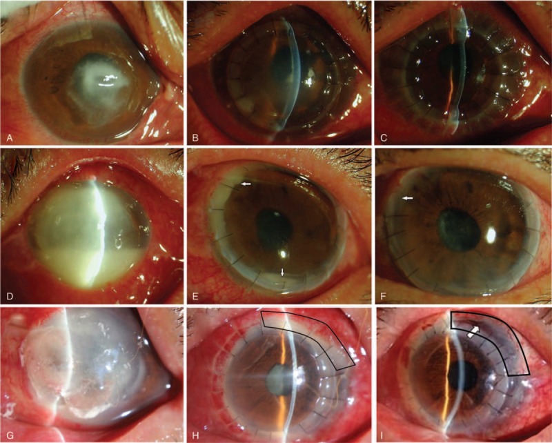
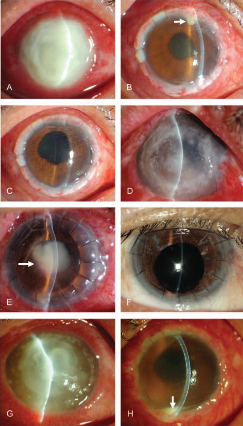
There are no standardized protocols or guidelines for the treatment of recurrent fungal keratitis after therapeutic keratoplasty. This study aimed to investigate the incidence of recurrent fungal keratitis after the primary keratoplasty and the visual outcome and prognosis after intervention for the recurrence.This was a retrospective study. Patients with recurrent fungal keratitis after lamellar keratoplasty (LK) or penetrating keratoplasty (PK) were treated with different antifungal regimens at Shandong Eye Hospital and Qingdao Eye Hospital between Januray 2004 and December 2015. The operative techniques included PK, focal excision, tectonic keratoplasty with a patch graft, lensectomy and vitrectomy, and combined operation. Patients were followed at 1, 2, and 3 months, and then every 6 months after surgery for 2 years. Best corrected visual acuity was assessed and recurrence was recorded. Good prognosis was defined as the presence of visual acuity.Fungal keratitis recurred in 112 of 1448 patients (112/1448, 7.7%) treated initially with PK or LK. The good prognosis rates for different sites of recurrent fungal keratitis were: overall, 93 of 112 (83.0%); recipient bed, 64 of 69 (92.8%); anterior chamber, 14 of 14 (100%); posterior segment, 10 of 16 (62.5%); and atypical, 5 of 13 (38.5%). There was no significant difference in the timing of recurrence between the good and poor prognosis groups (P = .518). Recurrence rates were similar between patients with PK (8.6%) and those with LK (6.0%; P > .05), but the good prognosis rate in patients with post-LK recurrence (96.8%) was higher than that in patients with post-PK recurrence (77.8%, P = .017).Individualized treatment according to recurrent sites of fungal keratitis can achieve a good prognosis in most patients.


Conflict of interest statement

The authors report no conflicts of interest.

Figures

Figure 1
Figure 1
Three representative cases of recipient bed recurrence. (A–C) Case 1. Male, 68 years’ old. (A) Central corneal infiltration; a little pus can be seen in the anterior chamber. (B) After lamellar keratoplasty (LK), the fungi recurred in the central recipient bed, showing as gray-white infiltration. (C) Medical therapy was not effective after recurrence. The graft was transparent after penetrating keratoplasty (PK). (D–F) Case 2. Female, 51 years’ old. (D) She had extensive corneal infection and a large amount of pus in the anterior chamber. (E) The infection recurred in the recipient bed after PK, showing gray-white infiltration. Purulent adhesion was observed at the interface of the recipient bed and graft. A little pus was observed in the anterior chamber (white arrow). (F) After recipient bed lesion resection at the recurrence site, the infection was controlled, the graft was transparent (white arrow), and the aqueous humor in the anterior chamber was clear. (G–I) Case 3. Male, 61 years’ old. (G) A large range of cornea was involved, and the corneaoscleral limbus was affected at the nasal side. Local perforation was observed. (H) Local recipient bed recurrence after PK. Purulent adhesion was observed at the interface of recipient bed and graft (black frame), the aqueous humor was turbid, and pus was observed in the lower anterior chamber. (I) Medical therapy was not effective after recurrence. The central graft and patch (black frame) were transparent and the aqueous humor in the anterior chamber was clear after lesion resection and patch PK treatment.
Figure 2
Figure 2
Three representative cases of anterior chamber, posterior segment, and atypical recurrence. (A–C) Case 1. Male, 50 years’ old. (A) Full corneal fungal infection affecting the corneal limbus, with peripheral cornea autolysis and thinning. (B) After total corneal transplantation, mushroom-shaped purulent exudation was seen in the upper corner of the chamber (white arrow) and the aqueous humor was turbid. (C) After removal of the mushroom-shaped purulent exudation and anterior chamber injection of antifungal drugs, the corneal grafts were transparent and the aqueous humor in the anterior chamber was clear. (D–F) Case 2. Male, 56 years’ old. (D) The preoperative corneal fungal infection involved total cornea and full corneal layers. The conditions of the anterior chamber could not be seen. (E) Fungal recurrence in the posterior segment after penetrating keratoplasty (PK). A mass-like purulent exudation was observed in the pupil area (white arrow). (F) Reexamination at 1 year after lensectomy + vitrectomy showed that the corneal graft was transparent, and aqueous humor was clear. (G–H) Case 3. Male, 33 years’ old. (G) The cornea was widely infected, locally thinned, and there were a large number of satellite foci. The conditions of the anterior chamber could not be seen. (H) Atypical recurrence was observed after PK. The Hypopyon was observed in the anterior chamber (white arrow). The aqueous humor was obviously turbid. The corneal graft was transparent, and the exact recurrence lesions were not seen in the recipient bed and the graft. After recurrence, medical therapy was not effective. An exploratory operation was performed. During surgery, it was found that the upper and nasal sclera were eroded. Massive empyema was observed in the ciliary body; the vitreous body also showed empyema. There was intraocular infection, and the eyeballs could not be kept and were removed.

Similar articles

Cited by

References

    1. Whitcher JP, Srinivasan M, Upadhyay MP. Corneal blindness: a global perspective. Bull World Health Organ 2001;79:214–21. - PMC - PubMed
    1. Song X, Xie L, Tan X, et al. A multi-center, cross-sectional study on the burden of infectious keratitis in China. PLoS One 2014;9:e113843. - PMC - PubMed
    1. Cao J, Yang Y, Yang W, et al. Prevalence of infectious keratitis in Central China. BMC Ophthalmol 2014;14:43. - PMC - PubMed
    1. Sauer A, Letscher-Bru V, Speeg-Schatz C, et al. In vitro efficacy of antifungal treatment using riboflavin/UV-A (365 nm) combination and amphotericin B. Invest Ophthalmol Vis Sci 2010;51:3950–3. - PubMed
    1. Carrasco MA, Genesoni G. Treatment of severe fungal keratitis with subconjunctival amphotericin B. Cornea 2011;30:608–11. - PubMed

MeSH terms

Substances