

Evaluation of blood flow on optic nerve head after pattern scan and conventional laser panretinal photocoagulation
- PMID: 31192968
- PMCID: PMC6587595
- DOI: 10.1097/MD.0000000000016062
Evaluation of blood flow on optic nerve head after pattern scan and conventional laser panretinal photocoagulation
Abstract
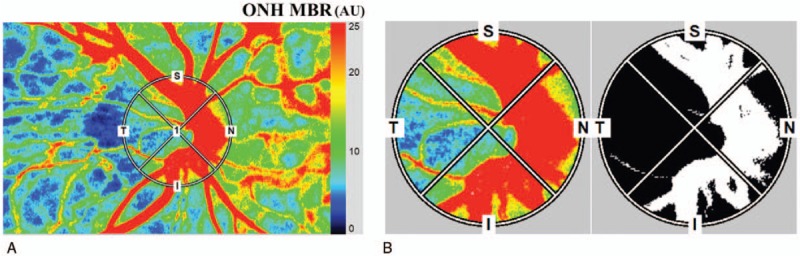
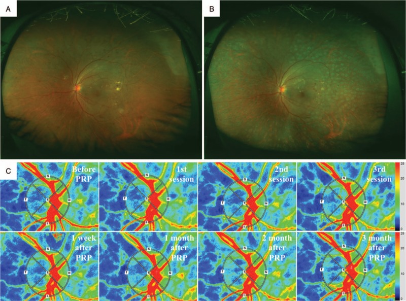
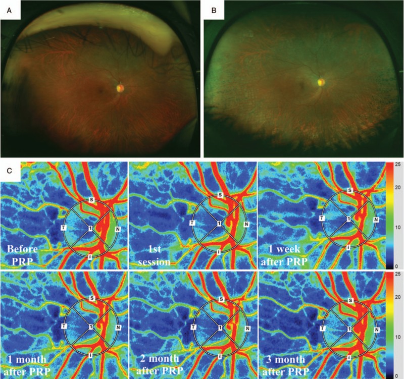
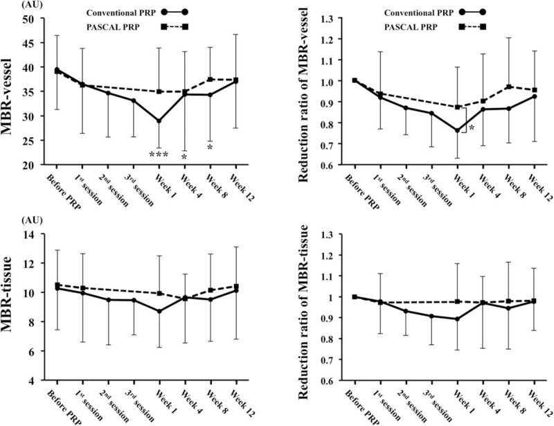
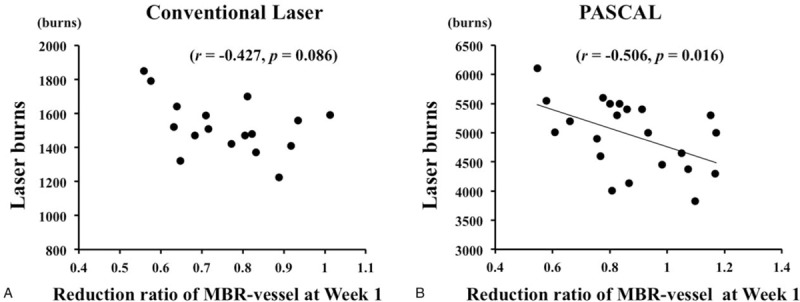
To evaluate the changes in the blood flow on retina and the optic nerve head (ONH) after conventional laser treatment and to compare it to that after patterned scanning laser (PASCAL) treatment in patients with severe nonproliferative diabetic retinopathy (S-NPDR).In this prospective, cross-sectional study, the blood flow on retina and the ONH was assessed by laser speckle flowgraphy using the mean blur rate (MBR) in 39 eyes with S-NPDR before, 1, 4, 8, 12 weeks after panretinal photocoagulation (PRP). Of 39 eyes, 17eyes with 17 patients treated by conventional laser and 22 eyes with 22 patients treated by PASCAL.The mean age was 55.5 ± 11.5 years in the conventional laser group, 55.6 ± 11.8 years in the PASCAL group. The MBR-vessel, which can be dominantly expressed as retinal blood flow, was significantly reduced after PRP treated by conventional laser (P < .001), but did not change after PRP treated by PASCAL. The ratio of MBR-vessel to the baseline was significantly lower in the conventional laser group only at Week 1 (P = .045). The MBR-tissue, which can be dominantly expressed as the ONH blood flow, did not significantly change after PRP in the both group. The multiple stepwise regression analysis revealed that the laser burns was an independent factor significantly correlated with the ratio of MBR-vessel at Week 1 to the baseline (β = -0.550, P = .012).The retinal blood flow was significantly reduced during the 12 weeks only after completion of PRP by conventional laser treatment. Our results indicate that short pulse on PRP treatment performed by the PASCAL would not significantly reduce the retinal blood flow.
Figures
Similar articles
-
Effects of photocoagulation on ocular blood flow in patients with severe non-proliferative diabetic retinopathy.PLoS One. 2017 Mar 29;12(3):e0174427. doi: 10.1371/journal.pone.0174427. eCollection 2017. PLoS One. 2017. PMID: 28355247 Free PMC article.
-
Effect of panretinal photocoagulation on retinal oxygen metabolism and ocular blood flow in diabetic retinopathy.Acta Ophthalmol. 2025 Jun;103(4):380-387. doi: 10.1111/aos.17442. Epub 2025 Jan 30. Acta Ophthalmol. 2025. PMID: 39887558
-
A randomized clinical trial evaluating choroidal blood flow and morphology after conventional and pattern scan laser panretinal photocoagulation.Sci Rep. 2018 Sep 20;8(1):14128. doi: 10.1038/s41598-018-32487-y. Sci Rep. 2018. PMID: 30237467 Free PMC article. Clinical Trial.
-
Pan retinal photocoagulation for proliferative diabetic retinopathy: pattern scan laser versus argon laser.Curr Opin Ophthalmol. 2014 May;25(3):164-70. doi: 10.1097/ICU.0000000000000048. Curr Opin Ophthalmol. 2014. PMID: 24663066 Review.
-
Comparison of Pain Scores Among Patients Undergoing Conventional and Novel Panretinal Photocoagulation for Diabetic Retinopathy: A Systematic Review.Clin Ophthalmol. 2021 Mar 2;15:953-971. doi: 10.2147/OPTH.S294227. eCollection 2021. Clin Ophthalmol. 2021. PMID: 33688163 Free PMC article. Review.
Cited by
-
Quantitative Evaluation of Fundus Autofluorescence in Laser Photocoagulation Scars for Diabetic Retinopathy: Conventional vs. Short-Pulse Laser.Life (Basel). 2023 Sep 12;13(9):1901. doi: 10.3390/life13091901. Life (Basel). 2023. PMID: 37763305 Free PMC article.
-
Effect of panretinal photocoagulation versus intravitreal bevacizumab injection on optic disc microcirculation in patients with diabetic retinopathy.Int J Retina Vitreous. 2024 Dec 18;10(1):98. doi: 10.1186/s40942-024-00621-w. Int J Retina Vitreous. 2024. PMID: 39695775 Free PMC article.
References
-
- The Diabetic Retinopathy Study Research Group. Preliminary report on effects of photocoagulation therapy. Am J Ophthalmol 1976;81:383–96. - PubMed
-
- The Diabetic Retinopathy Study Research Group. Photocoagulation treatment of proliferative diabetic retinopathy. Clinical application of Diabetic Retinopathy Study (DRS) findings, DRS Report Number 8. Ophthalmology 1981;88:583–600. - PubMed
-
- Early Treatment Diabetic Retinopathy Study Research Group. Early photocoagulation for diabetic retinopathy ETDRS report number 9. Ophthalmology 1991;98:766–85. - PubMed
-
- Wilkinson CP, Ferris FL, 3rd, Klein RE, et al. Proposed international clinical diabetic retinopathy and diabetic macular edema disease severity scales. Ophthalmology 2003;110:1677–82. - PubMed
Publication types
MeSH terms
LinkOut - more resources
Full Text Sources
Medical
Research Materials
