

Pancreatic schwannoma mimicking pancreatic cystadenoma: A case report and literature review of the imaging features
- PMID: 31192973
- PMCID: PMC6587594
- DOI: 10.1097/MD.0000000000016095
Pancreatic schwannoma mimicking pancreatic cystadenoma: A case report and literature review of the imaging features
Abstract
Introduction: Schwannomas, also known as neurilemmoma, are benign neoplasms that originating from Schwann cells in peripheral nerve sheaths. The head, neck, and extremities are the most common sites; however, pancreatic schwannomas are rare neoplasms. Accurate preoperative diagnosis of these tumors is very tough because of pancreatic schwannomas usually mimicking other cystic tumors. Here we present a case of pancreatic schwannoma misdiagnosed as pancreatic cystadenoma.
Patient concerns: We presented a rare case of a 55-year-old female admitted to our hospital for abdominal distension. The physical examination and results of laboratory testing reveal no abnormalities.
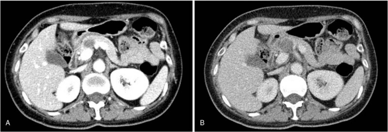
Diagnosis: A computed tomography (CT) scan detected a hypodense 2.4 cm × 2.6 cm mass with a clear margin at the neck of the pancreas. Pancreatic cystadenoma was strongly suspected.
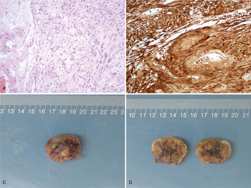
Interventions: The patient underwent robotic distal pancreatectomy with splenectomy. The gross specimen showed a pale and solid mass with a capsule.
Outcomes: Histological examination of the surgical specimen demonstrated a pancreatic schwannoma. Immunohistochemistry results were as follows: S-100 (+), CD117 (-), SMA (-), and Desmin (-). She was discharged on postoperative day 6 and no recurrence of the tumor happened during the 12-month follow-up.
Conclusion: Precise preoperative diagnosis of pancreatic schwannomas is very difficult despite the application of multiple imaging modalities. Surgery is the most effective treatment for this rare disease and the final diagnosis usually relies on pathology. Following complete tumor removal, patients with pancreatic schwannomas generally have a good prognosis.
Conflict of interest statement
The authors report no conflicts of interest.
Figures

References
-
- Bhattacharyya AK, Perrin R, Guha A. Peripheral nerve tumors: management strategies and molecular insights. J Neurooncol 2004;69:335–49. - PubMed
-
- Le Guellec S. [Nerve sheath tumours]. Ann Pathol 2015;35:54–70. - PubMed
-
- Akiyoshi T, Ueda Y, Yanai K, et al. Melanotic schwannoma of the pancreas: report of a case. Surg Today 2004;34:550–3. - PubMed
-
- von Dobschuetz E, Walch A, Werner M, et al. Giant ancient schwannoma of pancreatic head treated by extended pancreatoduodenectomy. Pancreatology 2004;4:505–8. - PubMed
Publication types
MeSH terms
LinkOut - more resources
Full Text Sources
Medical
