

Diagnostic challenges in congenital cytomegalovirus infection in pregnancy: A case report
- PMID: 31192993
- PMCID: PMC6510697
- DOI: 10.1016/j.crwh.2019.e00119
Diagnostic challenges in congenital cytomegalovirus infection in pregnancy: A case report
Abstract
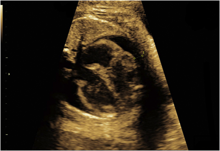
Cytomegalovirus is the most common congenital viral infection. Infection can cause developmental delay, sensorineural deafness and fetal death. Fetal damage is more severe when infection occurs in the first trimester of pregnancy. Prenatal ultrasound findings may be cerebral, such as ventriculomegaly, microcephaly and periventricular leukomalacia, as well as non-cerebral, such as echogenic bowel, ascites and pericardial effusion. We present a case of congenital cytomegalovirus infection in which the only ultrasound sign noted at routine second-trimester scan was low-grade echogenic bowel, a soft marker, which progressed to severe disease in the third trimester, when further investigation was prompted, leading to the diagnosis. Patients need to be counselled regarding the possible perinatal prognosis. Ultrasound markers can often but not always predict severity and, hence, counselling can be a challenge. Conclusion: A meticulous anatomy survey in mid-trimester remains the norm and ultrasound soft markers should prompt comprehensive testing for viral infections in pregnancy.
Keywords: Congenital cytomegalovirus infection; Fetal hydrops; Fetal pericardial effusion; IgG avidity.
Figures


Similar articles
-
Progressive lesions of central nervous system in microcephalic fetuses with suspected congenital Zika virus syndrome.Ultrasound Obstet Gynecol. 2017 Dec;50(6):717-722. doi: 10.1002/uog.17303. Epub 2017 Nov 8. Ultrasound Obstet Gynecol. 2017. PMID: 27644020
-
Secondary cytomegalovirus infection can cause severe fetal sequelae despite maternal preconceptional immunity.Ultrasound Obstet Gynecol. 2008 Apr;31(4):417-20. doi: 10.1002/uog.5255. Ultrasound Obstet Gynecol. 2008. PMID: 18383476
-
Feasibility of predicting the outcome of fetal infection with cytomegalovirus at the time of prenatal diagnosis.Am J Obstet Gynecol. 2016 Sep;215(3):342.e1-9. doi: 10.1016/j.ajog.2016.03.052. Epub 2016 Apr 8. Am J Obstet Gynecol. 2016. PMID: 27063062
-
Congenital cytomegalovirus infection in pregnancy: a review of prevalence, clinical features, diagnosis and prevention.Aust N Z J Obstet Gynaecol. 2016 Feb;56(1):9-18. doi: 10.1111/ajo.12408. Epub 2015 Sep 22. Aust N Z J Obstet Gynaecol. 2016. PMID: 26391432 Review.
-
[Ultrasound in the evaluation of intrauterine infection during pregnancy].Harefuah. 2009 Jul;148(7):460-4, 474. Harefuah. 2009. PMID: 19848336 Review. Hebrew.
Cited by
-
Multiple Cerebral Abnormalities at Third-trimester Ultrasound Scan in an Uncomplicated Pregnancy.J Med Ultrasound. 2021 Jul 24;31(1):69-71. doi: 10.4103/JMU.JMU_63_21. eCollection 2023 Jan-Mar. J Med Ultrasound. 2021. PMID: 37180622 Free PMC article. No abstract available.
-
Pathophysiology of Hyperechogenic Bowel in Congenitally Human Cytomegalovirus Infected Fetuses.Microorganisms. 2020 May 22;8(5):779. doi: 10.3390/microorganisms8050779. Microorganisms. 2020. PMID: 32455864 Free PMC article.
-
Investigation of Pre- and Postnatal Abnormalities Caused by Prenatal CMV Infection-Systematic Review.Children (Basel). 2025 May 6;12(5):607. doi: 10.3390/children12050607. Children (Basel). 2025. PMID: 40426786 Free PMC article. Review.
-
An Overview of Cytomegalovirus Infection in Pregnancy.Diagnostics (Basel). 2022 Oct 7;12(10):2429. doi: 10.3390/diagnostics12102429. Diagnostics (Basel). 2022. PMID: 36292118 Free PMC article. Review.
References
-
- Gaytant M.A., Steegers E.A., Semmekrot B.A., Merkus H.M., Galama J.M. Congenital cytomegalovirus infection: review of the epidemiology and outcome. Obstet. Gynecol. Surv. 2002;57:245–256. (Level III) - PubMed
-
- Alford C.A., Stagno S., Pass R.F., Britt W.J. Congenital and perinatal cytomegalovirus infections. Rev. Infect. Dis. 1990;12(Suppl. 7):S745–S753. - PubMed
-
- Kenneson A., Cannon M.J. Review and meta-analysis of the epidemiology of congenital cytomegalovirus (CMV) infection. Rev. Med. Virol. 2007;17:253. - PubMed
-
- McMullan B., Palasanthiran P., Jones C. Congenital cytomegalovirus - time to diagnosis, management and clinical sequelae in Australia: opportunities for earlier identification. Med. J. Aust. 2011;194:625–629. - PubMed
-
- Dahle A., Fowler K., Wright J. Longitudinal investigation of hearing disorders in children with congenital cytomegalovirus. J. Am. Acad. Audiol. 2000;11:283–290. - PubMed
Publication types
LinkOut - more resources
Full Text Sources
