. 2019 Apr 24:20:9-21.
doi: 10.1016/j.jare.2019.04.006. eCollection 2019 Nov.

LW6 enhances chemosensitivity to gemcitabine and inhibits autophagic flux in pancreatic cancer

Affiliations

LW6 enhances chemosensitivity to gemcitabine and inhibits autophagic flux in pancreatic cancer

Xianbin Zhang et al. J Adv Res. .

Abstract

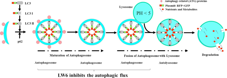
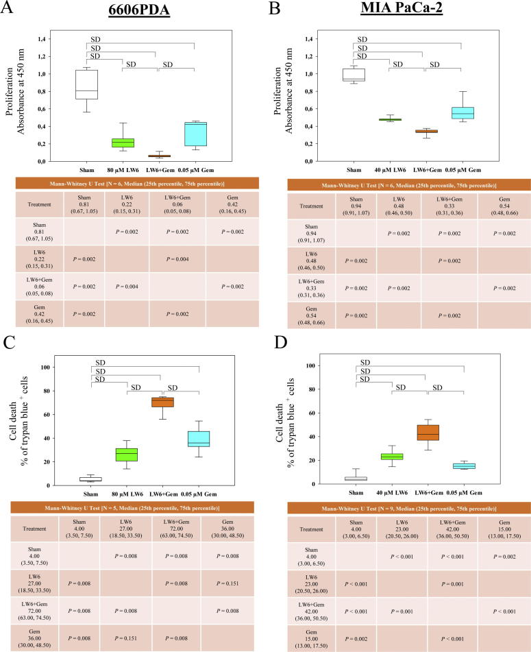
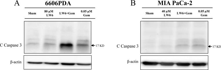
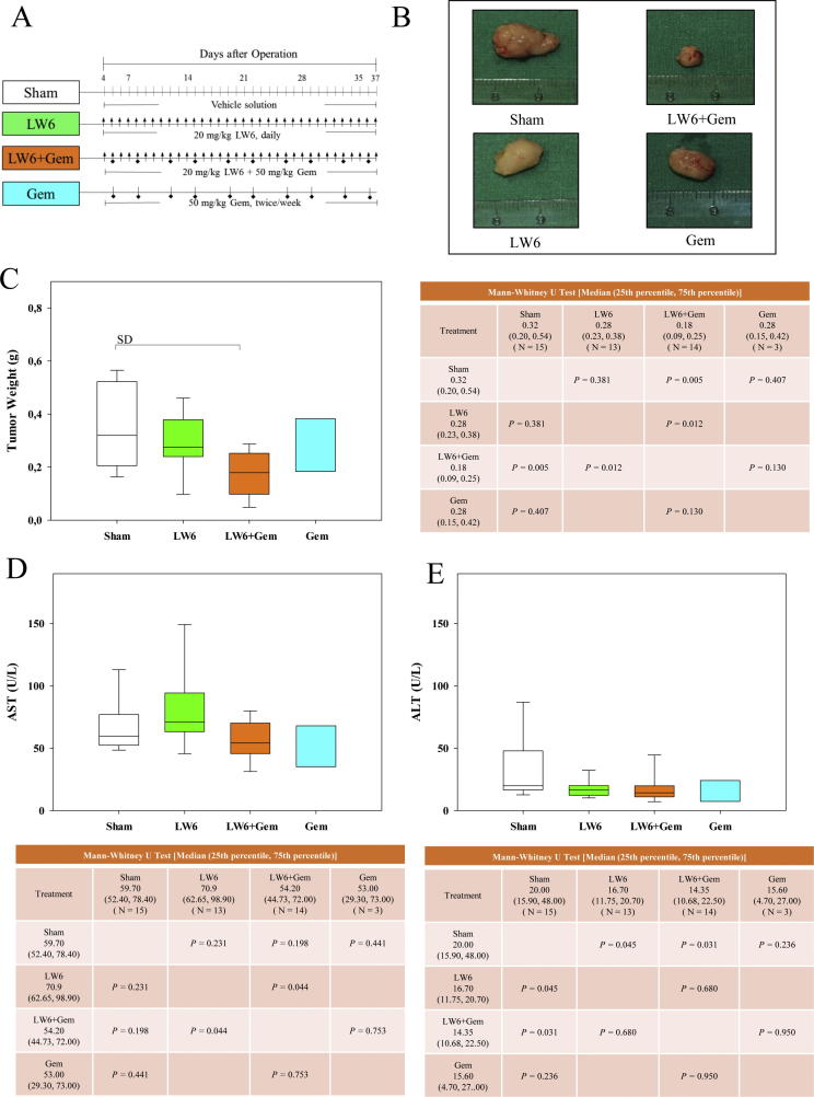
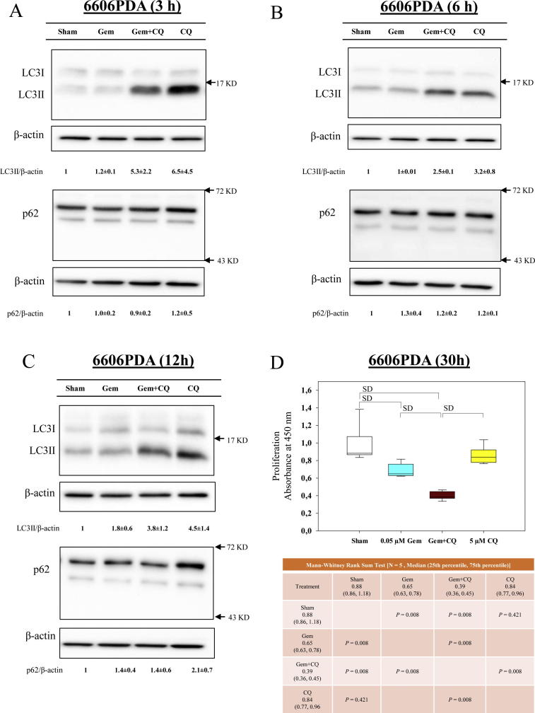
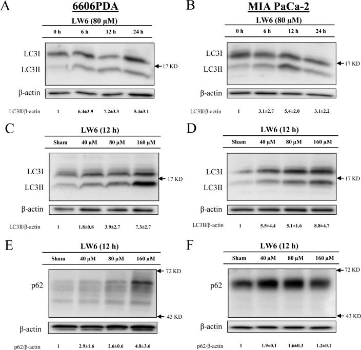
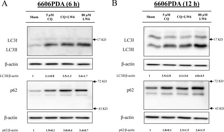
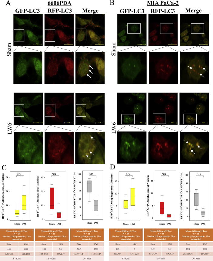
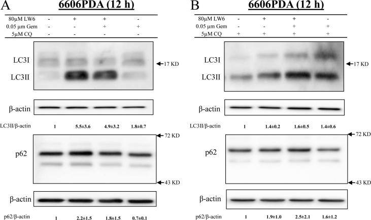
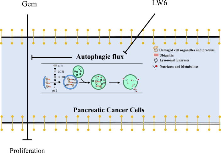
The efficacy of gemcitabine therapy is often insufficient for the treatment of pancreatic cancer. The current study demonstrated that LW6, a chemical inhibitor of hypoxia-inducible factor 1α, is a promising drug for enhancing the chemosensitivity to gemcitabine. LW6 monotherapy and the combination therapy of LW6 plus gemcitabine significantly inhibited cell proliferation and enhanced cell death in pancreatic cancer cells. This combination therapy also significantly reduced the tumor weight in a syngeneic orthotopic pancreatic carcinoma model without causing toxic side effects. In addition, this study provides insight into the mechanism of how LW6 interferes with the pathophysiology of pancreatic cancer. The results revealed that LW6 inhibited autophagic flux, which is defined by the accumulation of microtubule-associated protein 1 light chain 3 (LC3) and p62/SQSTM1. Moreover, these results were verified by the analysis of a tandem RFP-GFP-tagged LC3 protein. Thence, for the first time, these data demonstrate that LW6 enhances the anti-tumor effects of gemcitabine and inhibits autophagic flux. This suggests that the combination therapy of LW6 plus gemcitabine may be a novel therapeutic strategy for pancreatic cancer patients.

Keywords: 3-MA, 3-methyladenine; ATCC, American Type Culture Collection; Autophagy; BrdU, 5-bromo-2′-deoxyuridine; CQ, chloroquine; Combination therapy; Gemcitabine; LC3, microtubule-associated protein 1 light chain 3; LW6; Pancreatic cancer; p62, p62/SQSTM1.


Figures

None
Graphical abstract
Fig. 1
Fig. 1
LW6 impairs pancreatic cancer cells under normoxic and hypoxic conditions. 6606PDA (A) and MIAPaCa-2 (B) cells were treated with the indicated concentrations of LW6 and were cultured under normoxic and hypoxic conditions for 30 h. The inhibition of cell proliferation by LW6 was not influenced by hypoxic conditions. P < 0.01 indicates a significant difference.
Fig. 2
Fig. 2
LW6 inhibits proliferation and induces cell death. The amount of BrdU incorporation was analyzed after treating 6606PDA (A) and MIA PaCa-2 (B) cells with the indicated concentrations of LW6 for 30 h. In addition, 80 µM and 160 µM LW6 significantly inhibited cell proliferation compared to the cell proliferation of the Sham-treated cells or of the 40 µM LW6-treated cells (A and B). The percentage of dead cells was determined by a trypan blue assay after treating 6606PDA (C) and MIA PaCa-2 (D) cells with the indicated concentrations of LW6 for 48 h. The above mentioned concentrations of LW6 also significantly induced cell death compared to that in the Sham-treated cells or that in the 40 µM LW6-treated cells (C and D). P ≤ 0.008 indicates a significant difference (SD).
Fig. 3
Fig. 3
LW6 in combination with gemcitabine (Gem) inhibits proliferation and induces cell death. The amount of BrdU incorporation was analyzed after treating 6606PDA (A) and MIA PaCa-2 (B) cells with the indicated concentrations of LW6 or 0.05 µM gemcitabine for 30 h. The combinational therapy, 80 µM LW6 plus 0.05 µM gemcitabine, significantly inhibited the proliferation of 6606PDA cells compared to the proliferation of the Sham-treated cells or that of the monotherapy-treated cells (A). Similar results were obtained after treating MIA PaCa-2 cells with 40 µM LW6 plus 0.05 µM gemcitabine (B). The percentage of dead cells was determined by a trypan blue assay after treating 6606PDA (C) and MIA PaCa-2 (D) cells with the indicated concentrations of LW6 or 0.05 µM gemcitabine for 48 h. LW6 plus gemcitabine treatment also significantly induced cell death compared to the levels of cell death induced in the Sham-treated or the monotherapy-treated cells (C and D). P ≤ 0.008 indicates a significant difference (SD).
Fig. 4
Fig. 4
Gemcitabine (Gem) increases the accumulation of cleaved caspase 3 (C Caspase 3). Western blots were performed after treating 6606PDA (A) and MIA PaCa-2 (B) cells with gemcitabine, LW6, or the combined treatment for 54 h. Gemcitabine increased the accumulation of cleaved caspase 3 in both 6606PDA (A) and MIA PaCa-2 (B) cells compared with that in Sham-treated cells. A similar result was obtained after treating 6606PDA cells with LW6 (A). However, LW6 failed to increase the level of cleaved caspase 3 in MIA PaCa-2 cells (B). The experiment was repeated three times.
Fig. 5
Fig. 5
LW6 in combination with gemcitabine (Gem) decreases the tumor weight. Cohorts of mice were treated i.p. with the vehicle solution (symbol: line), 20 mg/kg LW6 (symbol: arrow), 50 mg/kg gemcitabine (symbol: square), or the combined treatment (symbol: arrow plus square) as indicated in the experimental schema (A). The treatment of mice with 20 mg/kg LW6 plus 50 mg/kg gemcitabine led to an obvious decrease in the tumor size (B). This combination therapy significantly reduced the tumor weight compared to the tumor weight in the Sham-treated mice (C). All indicated therapies did not induce liver toxicity as defined by aspartate transaminase (AST) activity (D) or alanine aminotransferase (ALT) activity (E) in the blood plasma. P ≤ 0.008 indicates a significant difference (SD).
Fig. 6
Fig. 6
Blocking autophagic flux enhances the anti-proliferative effect of gemcitabine (Gem). After treating 6606PDA cells by 0.05 µM gemcitabine for 3 h (A), 6 h (B) and 12 h (C), gemcitabine only had minor effects on the accumulation of LC3II and p62. However, blocking autophagy with 5 µM chloroquine (CQ) could enhance the anti-proliferation effect of gemcitabine (D). P ≤ 0.008 indicates a significant difference (SD).
Fig. 7
Fig. 7
LW6 regulates the accumulation of proteins involved in autophagy. After treating the cells with 80 µM LW6 or vehicle control (Sham) for the indicated time periods, the level of LC3II was increased in 6606PDA (A) and MIA PaCa-2 (B) cells. Treatment with LW6 for 12 h induced the accumulation of LC3II in a dose-dependent manner in both cell lines (C and D). In addition, this treatment also induced the accumulation of p62 in 6606PDA (E) and MIA PaCa-2 (F) cells. All experiments were repeated three times.
Fig. 8
Fig. 8
LW6 blocks autophagic flux. Treatment for 6 h (A) or 12 h (B) with 80 µM LW6 and during the last 6 h with 5 µM chloroquine (CQ) or a combination of both drugs caused accumulation of LC3II and p62. Compared to the LW6-treated cells, the combination therapy failed to increase the accumulation of LC3II and p62. All experiments were repeated three times.
Fig. 9
Fig. 9
LW6 increases the number of autophagosomes and decreases the number of autolysosomes. 6606PDA (A) and MIA PaCa-2 (B) cells were treated with 160 µM LW6 and 80 µM LW6 for 12 h, respectively. LW6 significantly increased the number of autophagosomes (C and D). In addition, LW6 also significantly decreased the number and percentage of autolysosomes in both cancer cell lines (C and D). The scale bar = 20 µm, P ≤ 0.05 indicates a significant difference (SD).
Fig. 10
Fig. 10
LW6 inhibits the autophagic process in gemcitabine-treated cells. LW6 plus gemcitabine caused a major increase in the accumulation of LC3II and p62 in the absence of CQ (A). Only a minor increase was observed when treating the cells with CQ for the last 6 h (B). All experiments were repeated three times.
Fig. 11
Fig. 11
Summary. The present study demonstrates that LW6 enhances the chemosensitivity to gemcitabine and inhibits autophagic flux in pancreatic cancer.

References

    1. Hu X.C., Zhang J., Xu B.H., Cai L., Ragaz J., Wang Z.H. Cisplatin plus gemcitabine versus paclitaxel plus gemcitabine as first-line therapy for metastatic triple-negative breast cancer (CBCSG006): a randomised, open-label, multicentre, phase 3 trial. Lancet Oncol. 2015;16:436–446. - PubMed
    1. Thatcher N., Hirsch F.R., Luft A.V., Szczesna A., Ciuleanu T.E., Dediu M. Necitumumab plus gemcitabine and cisplatin versus gemcitabine and cisplatin alone as first-line therapy in patients with stage IV squamous non-small-cell lung cancer (SQUIRE): an open-label, randomised, controlled phase 3 trial. Lancet Oncol. 2015;16:763–774. - PubMed
    1. Von Hoff D.D., Ervin T., Arena F.P., Chiorean E.G., Infante J., Moore M. Increased survival in pancreatic cancer with nab-paclitaxel plus gemcitabine. N Engl J Med. 2013;369:1691–1703. - PMC - PubMed
    1. Siegel R.L., Miller K.D., Jemal A. Cancer statistics, 2018. CA Cancer J Clin. 2018;68:7–30. - PubMed
    1. Lee K.1, Kang J.E., Park S.K., Jin Y., Chung K.S., Kim H.M. LW6, a novel HIF-1 inhibitor, promotes proteasomal degradation of HIF-1alpha via upregulation of VHL in a colon cancer cell line. Biochem Pharmacol. 2010;80:982–989. - PubMed