

IL-7R blockade reduces post-myocardial infarction-induced atherosclerotic plaque inflammation in ApoE-/- mice
- PMID: 31193072
- PMCID: PMC6517313
- DOI: 10.1016/j.bbrep.2019.100647
IL-7R blockade reduces post-myocardial infarction-induced atherosclerotic plaque inflammation in ApoE-/- mice
Erratum in
-
Erratum regarding missing Declaration of Competing Interest statements in previously published articles.Biochem Biophys Rep. 2021 Jan 7;25:100901. doi: 10.1016/j.bbrep.2020.100901. eCollection 2021 Mar. Biochem Biophys Rep. 2021. PMID: 33614995 Free PMC article.
Abstract
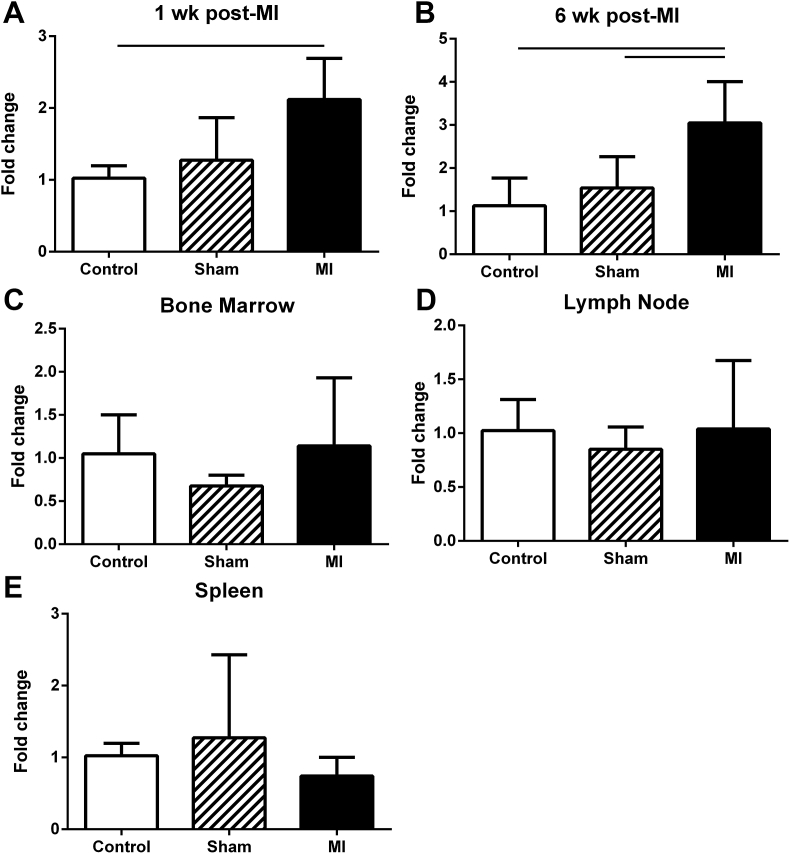
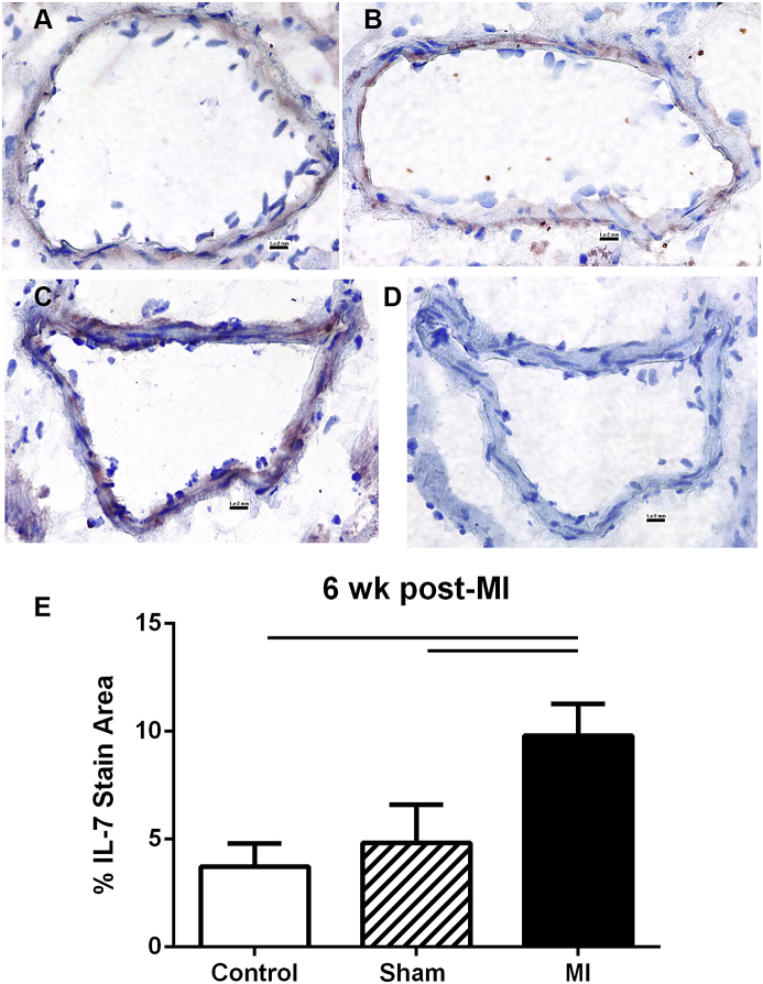
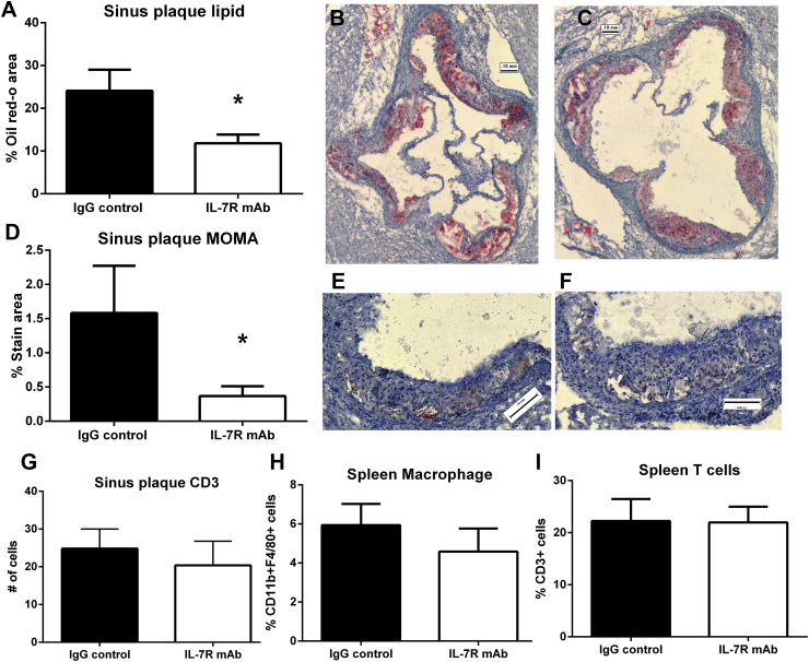
Modulating inflammation by targeting IL-1β reduces recurrent athero-thrombotic cardiovascular events without lipid lowering. This presents an opportunity to explore other pathways associated with the IL-1β signaling cascade to modulate the inflammatory response post-myocardial infarction (MI). IL-7 is a mediator of the inflammatory pathway involved in monocyte trafficking into atherosclerotic plaques and levels of IL-7 have been shown to be elevated in patients with acute MI. Recurrent athero-thrombotic events are believed to be mediated in part by index MI-induced exacerbation of inflammation in atherosclerotic plaques. The objective of the study was to assess the feasibility of IL-7R blockade to modulate atherosclerotic plaque inflammation following acute MI in ApoE-/- mice. Mice were fed Western diet for 12 weeks and then subjected to coronary occlusion to induce an acute MI. IL-7 expression was determined using qRT-PCR and immuno-staining, and IL-7R was assessed using flow cytometry. Plaque inflammation was evaluated using immunohistochemistry. IL-7R blockade was accomplished with monoclonal antibody to IL-7R. IL-7 mRNA expression was significantly increased in the cardiac tissue of mice subjected to MI but not in controls. IL-7 staining was observed in the coronary artery. Plaque macrophage and lipid content were significantly increased after MI. IL-7R antibody treatment but not control IgG significantly reduced macrophage and lipid content in atherosclerotic plaques. The results show that IL-7R antibody treatment reduces monocyte/macrophage and lipid content in the atherosclerotic plaque following MI suggesting a potential new target to mitigate increased plaque inflammation post-MI.
Keywords: IL-7R; Myocardial infarction; Plaque inflammation.
Figures
Similar articles
-
StemBell therapy stabilizes atherosclerotic plaques after myocardial infarction.Cytotherapy. 2018 Sep;20(9):1143-1154. doi: 10.1016/j.jcyt.2018.05.006. Epub 2018 Aug 12. Cytotherapy. 2018. PMID: 30107976
-
Lymphocytic myocarditis occurs with myocardial infarction and coincides with increased inflammation, hemorrhage and instability in coronary artery atherosclerotic plaques.Int J Cardiol. 2017 Apr 1;232:53-62. doi: 10.1016/j.ijcard.2017.01.052. Epub 2017 Jan 7. Int J Cardiol. 2017. PMID: 28087177
-
IL-1β inhibition partially negates the beneficial effects of diet-induced lipid lowering.bioRxiv [Preprint]. 2023 Oct 14:2023.10.13.562255. doi: 10.1101/2023.10.13.562255. bioRxiv. 2023. Update in: Arterioscler Thromb Vasc Biol. 2024 Jun;44(6):1379-1392. doi: 10.1161/ATVBAHA.124.320800. PMID: 37873280 Free PMC article. Updated. Preprint.
-
Recognition, pathophysiology, and management of acute myocardial infarction.Am J Health Syst Pharm. 2001 Sep 15;58(18):1709-18; quiz 1719-21. Am J Health Syst Pharm. 2001. PMID: 11571813 Review.
-
High-sensitivity C-reactive protein and atherosclerotic disease: from improved risk prediction to risk-guided therapy.Int J Cardiol. 2013 Oct 15;168(6):5126-34. doi: 10.1016/j.ijcard.2013.07.113. Epub 2013 Aug 24. Int J Cardiol. 2013. PMID: 23978367 Review.
Cited by
-
Cellular Heterogeneity of Activated Primary Human Macrophages and Associated Drug-Gene Networks: From Biology to Precision Therapeutics.Circulation. 2023 Nov 7;148(19):1459-1478. doi: 10.1161/CIRCULATIONAHA.123.064794. Epub 2023 Oct 18. Circulation. 2023. PMID: 37850387 Free PMC article.
-
The commonness in immune infiltration of rheumatoid arthritis and atherosclerosis: Screening for central targets via microarray data analysis.Front Immunol. 2022 Oct 13;13:1013531. doi: 10.3389/fimmu.2022.1013531. eCollection 2022. Front Immunol. 2022. PMID: 36311761 Free PMC article.
-
Haplo-insufficiency of Profilin1 in vascular endothelial cells is beneficial but not sufficient to confer protection against experimentally induced atherosclerosis.Cytoskeleton (Hoboken). 2025 Mar;82(3):81-90. doi: 10.1002/cm.21859. Epub 2024 Apr 16. Cytoskeleton (Hoboken). 2025. PMID: 38623956 Free PMC article.
-
The Identification and Evaluation of Interleukin-7 as a Myokine Biomarker for Peripheral Artery Disease Prognosis.J Clin Med. 2024 Jun 19;13(12):3583. doi: 10.3390/jcm13123583. J Clin Med. 2024. PMID: 38930112 Free PMC article.
-
Upregulated anti-angiogenic miR-424-5p in type 1 diabetes (model of subclinical cardiovascular disease) correlates with endothelial progenitor cells, CXCR1/2 and other parameters of vascular health.Stem Cell Res Ther. 2021 May 14;12(1):249. doi: 10.1186/s13287-021-02332-7. Stem Cell Res Ther. 2021. PMID: 33985567 Free PMC article.
References
-
- Kikkert W.J., Hoebers L.P., Damman P., Lieve K.V., Claessen B.E., Vis M.M., Baan J., Jr., Koch K.T., de Winter R.J., Piek J.J., Tijssen J.G., Henriques J.P. Recurrent myocardial infarction after primary percutaneous coronary intervention for ST-segment elevation myocardial infarction. Am. J. Cardiol. 2014;113(2):229–235. - PubMed
-
- Remskar M., Horvat M., Hojker S., Noc M. Procalcitonin in patients with acute myocardial infarction Wien. Klin. Wochenschr. 2002;114(5–6):205–210. - PubMed
-
- Joshi N.V., Toor I., Shah A.S., Carruthers K., Vesey A.T., Alam S.R., Sills A., Hoo T.Y., Melville A.J., Langlands S.P., Jenkins W.S., Uren N.G., Mills N.L., Fletcher A.M., van Beek E.J., Rudd J.H., Fox K.A., Dweck M.R., Newby D.E. Systemic atherosclerotic inflammation following acute myocardial infarction: myocardial infarction begets myocardial infarction. J. Am. Heart Assoc. 2015;4(9):e001956. - PMC - PubMed
-
- Ridker P.M., Everett B.M., Thuren T., MacFadyen J.G., Chang W.H., Ballantyne C., Fonseca F., Nicolau J., Koenig W., Anker S.D., Kastelein J.J.P., Cornel J.H., Pais P., Pella D., Genest J., Cifkova R., Lorenzatti A., Forster T., Kobalava Z., Vida-Simiti L., Flather M., Shimokawa H., Ogawa H., Dellborg M., Rossi P.R.F., Troquay R.P.T., Libby P., Glynn R.J. Antiinflammatory therapy with canakinumab for atherosclerotic disease. N. Engl. J. Med. 2017;377(12):1119–1131. - PubMed
LinkOut - more resources
Full Text Sources
Other Literature Sources
Miscellaneous
