

Predictive value of genetic testing for inherited retinal diseases in patients with suspected atypical autoimmune retinopathy
- PMID: 31193260
- PMCID: PMC6523031
- DOI: 10.1016/j.ajoc.2019.100461
Predictive value of genetic testing for inherited retinal diseases in patients with suspected atypical autoimmune retinopathy
Abstract
Purpose: The clinical features of autoimmune retinopathy (AIR) can resemble and be difficult to differentiate from inherited retinal degenerations (IRDs). Misdiagnosis of an IRD as AIR causes unnecessary treatment with immunosuppressive agents. The purpose of this study is to calculate the predictive value of genetic testing for IRDs in patients with suspected AIR and provide clinical examples where genetic testing has been useful.
Methods: We identified patients seen at MEEI between April 2013 and January 2017 for whom the differentiation of AIR vs. IRDs was difficult based on clinical assessment alone. All patients had some atypical features for AIR, but tested positive for anti-retinal antibodies. Within this group, we identified six patients who had genetic testing for IRDs with the Genetic Eye Disease panel for retinal genes (GEDi-R). We calculated the positive predictive value (PPV) and negative predictive value (NPV) of genetic testing in a population with approximately equal numbers of IRD and AIR patients.
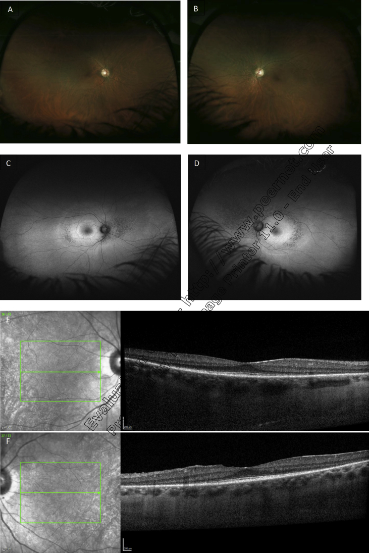
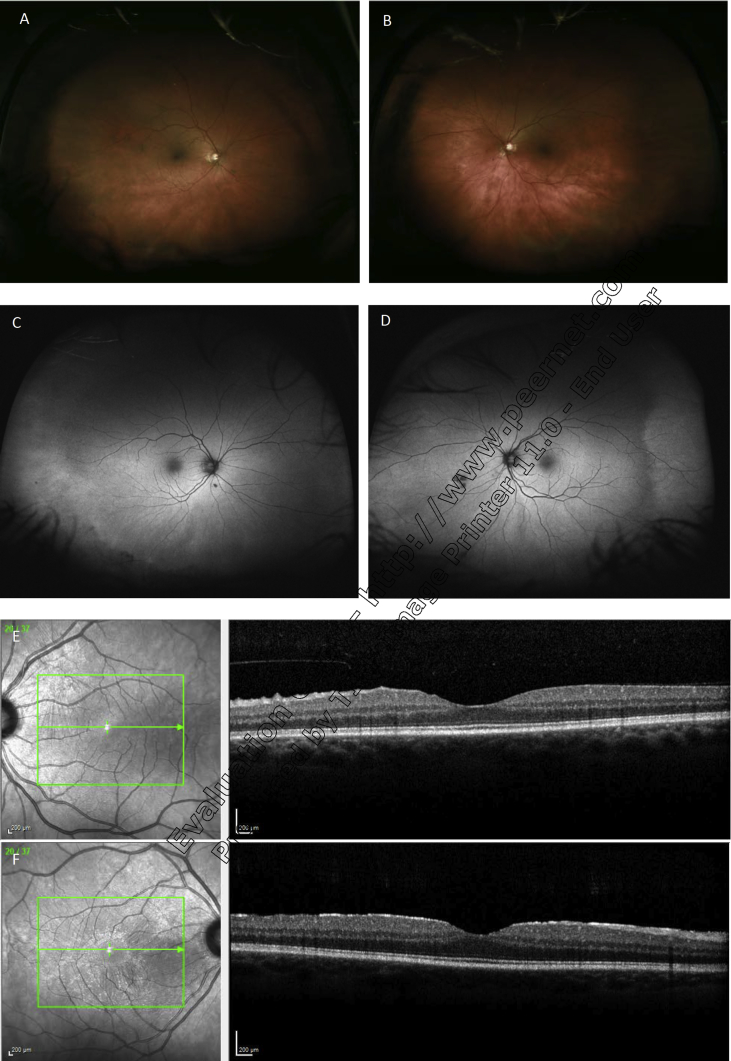
Results: Six patients had clinical features that made distinguishing between IRDs and AIR on a clinical basis difficult and were sent for genetic testing: four women and two men with a mean age of 59.5 years. In two of these six patients, genetic diagnoses were made based upon the identification of known pathogenic variants in the common IRD genes USH2A and RHO. Two patients had variants of unknown significance within genes associated with IRDs, and the other two had no relevant genetic findings. Given the 60% sensitivity and 3% false positive rate for GEDi-R testing and assuming a 50% pre-test probability of having an IRD, the PPV for GEDi-R for detecting IRD is 95.2% and the NPV is 70.8%.
Conclusions and importance: In patients for whom the differential diagnosis of AIR and IRDs is unclear based on clinical information, genetic testing can be a valuable tool when it identifies an IRD, sparing the patient unnecessary immunosuppressive treatment. However, the test has a low NPV so a negative genetic testing result does not confidently exclude IRD as the true diagnosis.
Keywords: Autoimmune retinopathy; Cancer associated-retinopathy; Genetic testing; Inherited retinal degeneration.
Figures
Similar articles
-
The Current State of Genetic Testing Platforms for Inherited Retinal Diseases.Ophthalmol Retina. 2022 Aug;6(8):702-710. doi: 10.1016/j.oret.2022.03.011. Epub 2022 Mar 18. Ophthalmol Retina. 2022. PMID: 35307606 Free PMC article.
-
DIAGNOSTIC YIELD OF AN INHERITED RETINAL DISEASE GENE PANEL IN RETINOPATHY OF UNKNOWN ORIGIN.Retina. 2024 Sep 1;44(9):1597-1607. doi: 10.1097/IAE.0000000000004155. Retina. 2024. PMID: 39167581
-
Current management of inherited retinal degenerations in Portugal (IRD-PT survey).Sci Rep. 2024 Sep 14;14(1):21473. doi: 10.1038/s41598-024-72589-4. Sci Rep. 2024. PMID: 39277603 Free PMC article.
-
The Impact of Inherited Retinal Diseases in the Republic of Ireland (ROI) and the United Kingdom (UK) from a Cost-of-Illness Perspective.Clin Ophthalmol. 2020 Mar 5;14:707-719. doi: 10.2147/OPTH.S241928. eCollection 2020. Clin Ophthalmol. 2020. PMID: 32184557 Free PMC article. Review.
-
Next-Generation Sequencing Applications for Inherited Retinal Diseases.Int J Mol Sci. 2021 May 26;22(11):5684. doi: 10.3390/ijms22115684. Int J Mol Sci. 2021. PMID: 34073611 Free PMC article. Review.
Cited by
-
Correlation of Immunological Markers with Disease and Clinical Outcome Measures in Patients with Autoimmune Retinopathy.Transl Vis Sci Technol. 2020 Jun 16;9(7):15. doi: 10.1167/tvst.9.7.15. eCollection 2020 Jun. Transl Vis Sci Technol. 2020. PMID: 32832222 Free PMC article.
-
Patient derived stem cells for discovery and validation of novel pathogenic variants in inherited retinal disease.Prog Retin Eye Res. 2021 Jul;83:100918. doi: 10.1016/j.preteyeres.2020.100918. Epub 2020 Oct 29. Prog Retin Eye Res. 2021. PMID: 33130253 Free PMC article. Review.
-
Higher throughput assays for understanding the pathogenicity of variants of unknown significance (VUS) in the RPE65 gene.bioRxiv [Preprint]. 2025 Feb 5:2025.01.31.635952. doi: 10.1101/2025.01.31.635952. bioRxiv. 2025. PMID: 39975398 Free PMC article. Preprint.
-
Using Molecular Diagnostics for Inherited Retinal Dystrophies: The 6 "I"s That Are Necessary to Diagnose 2 Eyes Genetically.Ophthalmol Sci. 2021 Mar 19;1(1):100018. doi: 10.1016/j.xops.2021.100018. eCollection 2021 Mar. Ophthalmol Sci. 2021. PMID: 36246004 Free PMC article. No abstract available.
-
Anti-TRPM1 autoantibody-positive unilateral melanoma associated retinopathy (MAR) triggered by immunotherapy recapitulates functional and structural details of TRPM1-associated congenital stationary night blindness.Am J Ophthalmol Case Rep. 2024 Jul 5;36:102098. doi: 10.1016/j.ajoc.2024.102098. eCollection 2024 Dec. Am J Ophthalmol Case Rep. 2024. PMID: 39109318 Free PMC article.
References
-
- Adamus G., Machnicki M., Elerding H. Antibodies to recoverin induce apoptosis of photoreceptor and bipolar cells in vivo. J Autoimmun. 1998;11(5):523–533. - PubMed
-
- Thirkill C.E., FitzGerald P., Sergott R.C. Cancer-associated retinopathy (CAR syndrome) with antibodies reacting with retinal, optic-nerve, and cancer cells. N Engl J Med. 1989;321(23):1589–1594. - PubMed
-
- Shimazaki K., Jirawuthiworavong G.V., Heckenlively J.R., Gordon L.K. Frequency of anti-retinal antibodies in normal human serum. J Neuro Ophthalmol. 2008;28(1):5–11. - PubMed
LinkOut - more resources
Full Text Sources
Miscellaneous
