

Differentiation of embryonic stem cells into inner ear vestibular hair cells using vestibular cell derived-conditioned medium
- PMID: 31193276
- PMCID: PMC6525281
- DOI: 10.1016/j.bbrep.2019.100649
Differentiation of embryonic stem cells into inner ear vestibular hair cells using vestibular cell derived-conditioned medium
Abstract
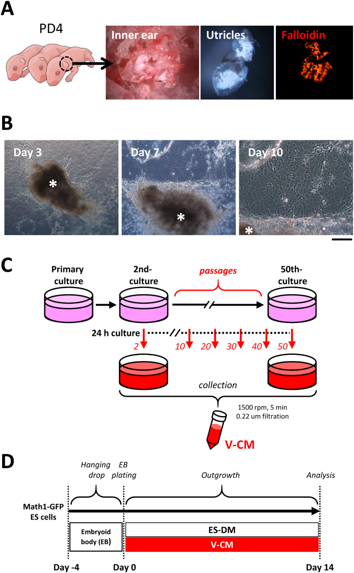
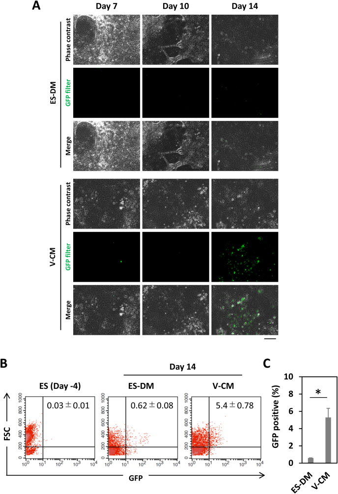
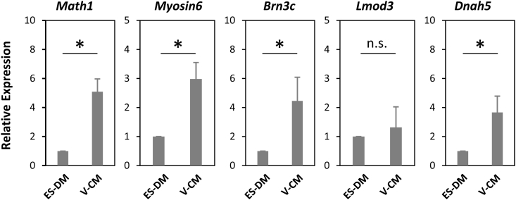
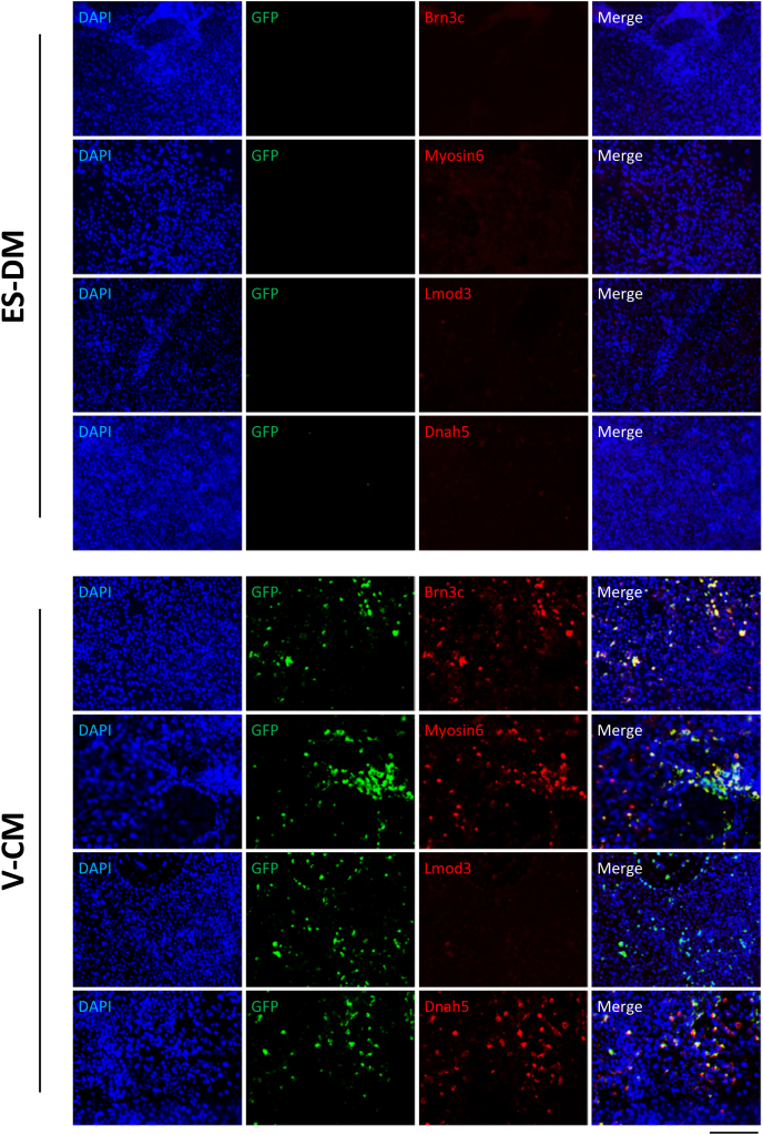
Vestibular hair cells (V-HCs) in the inner ear have important roles and various functions. When V-HCs are damaged, crippling symptoms, such as vertigo, visual field oscillation, and imbalance, are often seen. Recently, several studies have reported differentiation of embryonic stem (ES) cells, as pluripotent stem cells, to HCs, though a method for producing V-HCs has yet to be established. In the present study, we used vestibular cell conditioned medium (V-CM) and effectively induced ES cells to differentiate into V-HCs. Expressions of V-HC-related markers (Math1, Myosin6, Brn3c, Dnah5) were significantly increased in ES cells cultured in V-CM for 2 weeks, while those were not observed in ES cells cultured without V-CM. On the other hand, the cochlear HC-related marker Lmod3 was either not detected or detected only faintly in those cells when cultured in V-CM. Our results demonstrate that V-CM has an ability to specifically induce differentiation of ES cells into V-HCs.
Keywords: Conditioned medium; Differentiation; Embryonic stem cells; Hair cells; Inner ear; Vestibular.
Figures
Similar articles
-
Culture of organoids with vestibular cell-derived factors promotes differentiation of embryonic stem cells into inner ear vestibular hair cells.J Biosci Bioeng. 2023 Feb;135(2):143-150. doi: 10.1016/j.jbiosc.2022.11.005. Epub 2022 Dec 8. J Biosci Bioeng. 2023. PMID: 36503871
-
Efficient induction of inner ear hair cell-like cells from mouse ES cells using combination of Math1 transfection and conditioned medium from ST2 stromal cells.Stem Cell Res. 2017 Aug;23:50-56. doi: 10.1016/j.scr.2017.06.013. Epub 2017 Jul 1. Stem Cell Res. 2017. PMID: 28689068
-
In vitro differentiation of mouse embryonic stem cells into inner ear hair cell-like cells using stromal cell conditioned medium.Cell Death Dis. 2012 May 24;3(5):e314. doi: 10.1038/cddis.2012.56. Cell Death Dis. 2012. PMID: 22622133 Free PMC article.
-
Opportunities and limits of the one gene approach: the ability of Atoh1 to differentiate and maintain hair cells depends on the molecular context.Front Cell Neurosci. 2015 Feb 5;9:26. doi: 10.3389/fncel.2015.00026. eCollection 2015. Front Cell Neurosci. 2015. PMID: 25698932 Free PMC article. Review.
-
Stem cells for the replacement of inner ear neurons and hair cells.Int J Dev Biol. 2007;51(6-7):655-61. doi: 10.1387/ijdb.072372rm. Int J Dev Biol. 2007. PMID: 17891724 Review.
Cited by
-
Frailty of the.J Int Adv Otol. 2024 Jan;20(1):76-80. doi: 10.5152/iao.2024.231181. J Int Adv Otol. 2024. PMID: 38454293 Free PMC article.
-
Retinoic acid induces differentiation of cochlear neural progenitor cells into hair cells.Braz J Otorhinolaryngol. 2022 Nov-Dec;88(6):962-967. doi: 10.1016/j.bjorl.2021.01.005. Epub 2021 Feb 21. Braz J Otorhinolaryngol. 2022. PMID: 33707121 Free PMC article.
-
Differentiation of embryonic stem cells into a putative hair cell-progenitor cells via co-culture with HEI-OC1 cells.Sci Rep. 2021 Jul 6;11(1):13893. doi: 10.1038/s41598-021-93049-3. Sci Rep. 2021. PMID: 34230535 Free PMC article.
References
-
- Zine A. Molecular mechanisms that regulate auditory hair-cell differentiation in the mammalian cochlea. Mol. Neurobiol. 2003;27:223–238. - PubMed
-
- Wiest G. The origins of vestibular science. Ann. N. Y. Acad. Sci. 2015;1343:1–9. - PubMed
-
- Day B.L., Fitzpatrick R.C. The vestibular system. Curr. Biol. 2005;15:R583–R586. - PubMed
LinkOut - more resources
Full Text Sources
