

Biofunctionalization of metallic implants by calcium phosphate coatings
- PMID: 31193406
- PMCID: PMC6529680
- DOI: 10.1016/j.bioactmat.2019.05.001
Biofunctionalization of metallic implants by calcium phosphate coatings
Abstract
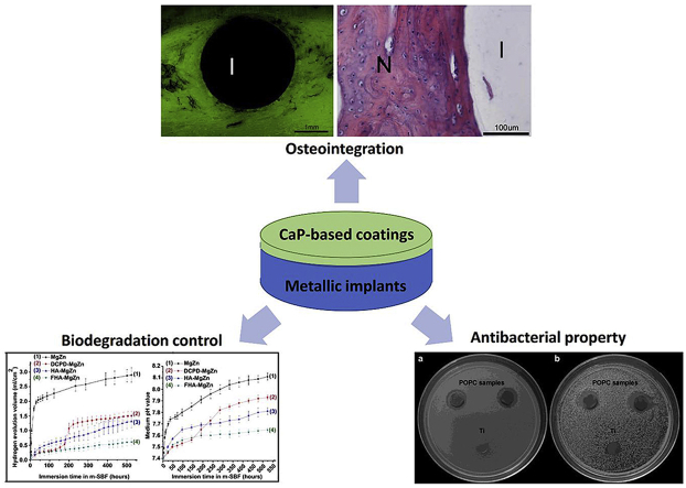
Metallic materials have been extensively applied in clinical practice due to their unique mechanical properties and durability. Recent years have witnessed broad interests and advances on surface functionalization of metallic implants for high-performance biofunctions. Calcium phosphates (CaPs) are the major inorganic component of bone tissues, and thus owning inherent biocompatibility and osseointegration properties. As such, they have been widely used in clinical orthopedics and dentistry. The new emergence of surface functionalization on metallic implants with CaP coatings shows promise for a combination of mechanical properties from metals and various biofunctions from CaPs. This review provides a brief summary of state-of-art of surface biofunctionalization on implantable metals by CaP coatings. We first glance over different types of CaPs with their coating methods and in vitro and in vivo performances, and then give insight into the representative biofunctions, i.e. osteointegration, corrosion resistance and biodegradation control, and antibacterial property, provided by CaP coatings for metallic implant materials.
Keywords: Biodegradation; Calcium phosphates; Metallic implant materials; Osteointegration; Surface biofunctionalization.
Figures
Similar articles
-
Advances in Multifunctional Bioactive Coatings for Metallic Bone Implants.Materials (Basel). 2022 Dec 25;16(1):183. doi: 10.3390/ma16010183. Materials (Basel). 2022. PMID: 36614523 Free PMC article. Review.
-
Tantalum-incorporated hydroxyapatite coating on titanium implants: its mechanical and in vitro osteogenic properties.J Mater Sci Mater Med. 2019 Oct 3;30(10):111. doi: 10.1007/s10856-019-6308-9. J Mater Sci Mater Med. 2019. PMID: 31583537
-
Investigation of duty cycle effect on corrosion properties of electrodeposited calcium phosphate coatings.Mater Sci Eng C Mater Biol Appl. 2016 Nov 1;68:681-686. doi: 10.1016/j.msec.2016.06.010. Epub 2016 Jun 4. Mater Sci Eng C Mater Biol Appl. 2016. PMID: 27524068
-
Calcium Phosphate Bioceramics: A Review of Their History, Structure, Properties, Coating Technologies and Biomedical Applications.Materials (Basel). 2017 Mar 24;10(4):334. doi: 10.3390/ma10040334. Materials (Basel). 2017. PMID: 28772697 Free PMC article. Review.
-
Corrosion Resistance and Cytocompatibility of Magnesium-Calcium Alloys Modified with Zinc- or Gallium-Doped Calcium Phosphate Coatings.ACS Appl Mater Interfaces. 2022 Jan 12;14(1):104-122. doi: 10.1021/acsami.1c16307. Epub 2021 Dec 27. ACS Appl Mater Interfaces. 2022. PMID: 34958199
Cited by
-
Self-adhesive lubricated coating for enhanced bacterial resistance.Bioact Mater. 2021 Feb 4;6(8):2535-2545. doi: 10.1016/j.bioactmat.2021.01.028. eCollection 2021 Aug. Bioact Mater. 2021. PMID: 33615044 Free PMC article.
-
Insights into self-healing behavior and mechanism of dicalcium phosphate dihydrate coating on biomedical Mg.Bioact Mater. 2020 Aug 12;6(1):158-168. doi: 10.1016/j.bioactmat.2020.07.019. eCollection 2021 Jan. Bioact Mater. 2020. PMID: 32817922 Free PMC article.
-
Assessing the osseointegration potential of a strontium releasing nanostructured titanium oxide surface: A biomechanical study in the rabbit tibia plateau model.Clin Exp Dent Res. 2024 Feb;10(1):e812. doi: 10.1002/cre2.812. Epub 2023 Dec 3. Clin Exp Dent Res. 2024. PMID: 38044566 Free PMC article.
-
In Vitro Study of Degradation and Cytocompatibility of Ceramics/PLA Composite Coating on Pure Zinc for Orthopedic Application.Front Bioeng Biotechnol. 2022 Mar 4;10:856986. doi: 10.3389/fbioe.2022.856986. eCollection 2022. Front Bioeng Biotechnol. 2022. PMID: 35309984 Free PMC article.
-
Iodine-Loaded Calcium Titanate for Bone Repair with Sustainable Antibacterial Activity Prepared by Solution and Heat Treatment.Nanomaterials (Basel). 2021 Aug 26;11(9):2199. doi: 10.3390/nano11092199. Nanomaterials (Basel). 2021. PMID: 34578515 Free PMC article.
References
-
- Crubezy E. False teeth of the Roman world. Nature. 1998;6662(391):29. - PubMed
-
- Park J.B., Lakes R.S., editors. Biomaterials. Springer New York; New York, NY: 2007. Metallic implant materials; pp. 99–137.
-
- Niinomi M. Development of new metallic alloys for biomedical applications. Acta Biomater. 2012;11(8):3888–3903. - PubMed
-
- Zheng Y.F. 2014. Biodegradable Metals, Materials Science and Engineering: R: Reports77; pp. 1–34.
Publication types
Grants and funding
LinkOut - more resources
Full Text Sources
Research Materials
Miscellaneous
