

The full recovery of mice (Mus Musculus C57BL/6 strain) from virus-induced sarcoma after treatment with a complex of DDMC delivery system and sncRNAs
- PMID: 31193489
- PMCID: PMC6531865
- DOI: 10.1016/j.ncrna.2019.03.001
The full recovery of mice (Mus Musculus C57BL/6 strain) from virus-induced sarcoma after treatment with a complex of DDMC delivery system and sncRNAs
Abstract
Background: Virus-induced cellular genetic modifications result in the development of many human cancers.
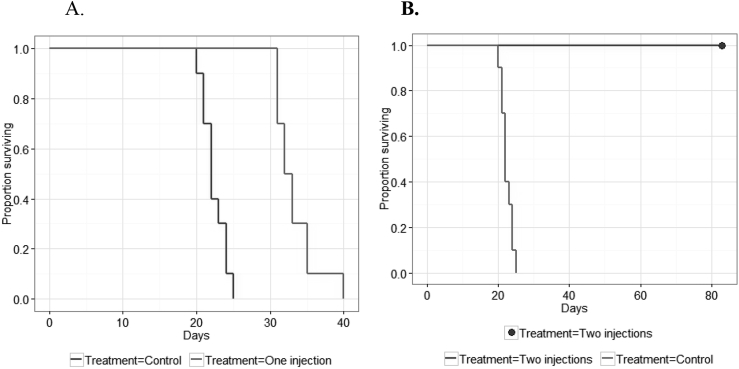
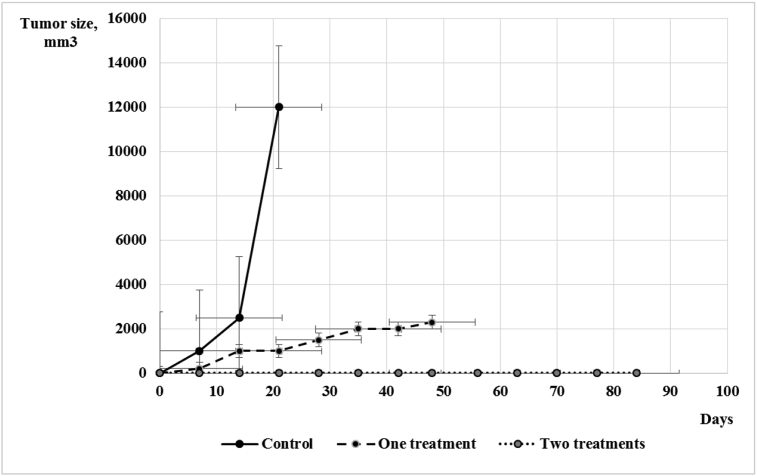
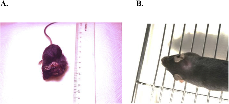
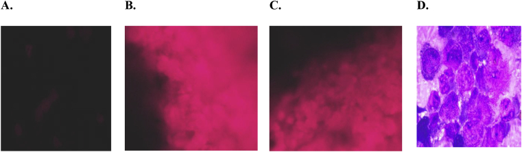
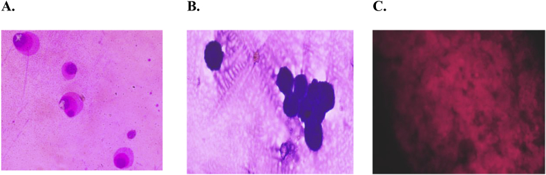
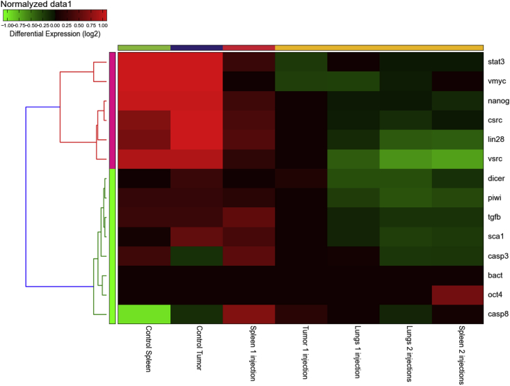
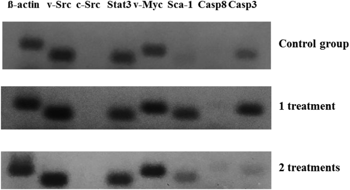
Methods: In our experiments, we used the RVP3 cell line, which produce primary mouse virus-induced sarcoma in 100% of cases. Inbreed 4-week-old female C57BL/6 mice were injected subcutaneously in the interscapular region with RVP3 cells. Three groups of mice were used. For treatment, one and/or two intravenous injections of a complex of small non-coding RNAs (sncRNAs) a-miR-155, piR-30074, and miR-125b with a 2-diethylaminoethyl-dextran methyl methacrylate copolymer (DDMC) delivery system were used. The first group consisted of untreated animals (control). The second group was treated with one injection of complex DDMC/sncRNAs (1st group). The third group was treated with two injections of complex DDMC/sncRNAs (2nd group). The tumors were removed aseptically, freed of necrotic material, and used with spleen and lungs for subsequent RT-PCR and immunofluorescence experiments, or stained with Leishman-Romanowski dye.
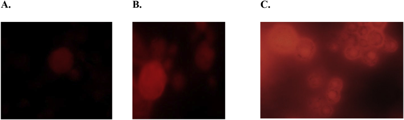
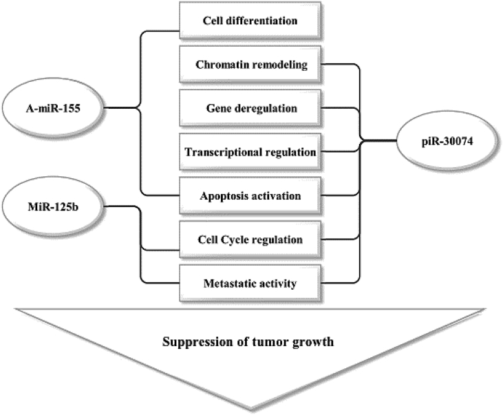
Results: As a result, the mice fully recovered from virus-induced sarcoma after two treatments with a complex including the DDMC vector and a-miR-155, piR-30074, and miR-125b. In vitro studies showed genetic and morphological transformations of murine cancer cells after the injections.
Conclusions: Treatment of virus-induced sarcoma of mice with a-miR-155, piR-30074, and miR-125b as active component of anti-cancer complex and DDMC vector as delivery system due to epigenetic-regulated transformation of cancer cells into cells with non-cancerous physiology and morphology and full recovery of disease.
Keywords: DDMC vector; Epigenetic therapy; Mice; Sarcoma; Small non-coding RNAs; Src tyrosine kinase.
Figures
Similar articles
-
DDMC-p53 gene therapy with or without cisplatin and microwave ablation.Onco Targets Ther. 2015 May 20;8:1165-73. doi: 10.2147/OTT.S83794. eCollection 2015. Onco Targets Ther. 2015. PMID: 26056480 Free PMC article.
-
Medicinal facilities to B16F10 melanoma cells for distant metastasis control with a supramolecular complex by DEAE-dextran-MMA copolymer/paclitaxel.Drug Deliv Transl Res. 2015 Feb;5(1):38-50. doi: 10.1007/s13346-014-0213-z. Drug Deliv Transl Res. 2015. PMID: 25787338
-
Characteristics of DEAE-dextran-MMA graft copolymer as a nonviral gene carrier.Nanomedicine. 2007 Sep;3(3):184-91. doi: 10.1016/j.nano.2007.07.002. Nanomedicine. 2007. PMID: 17765639
-
Anticancer efficacy of a supramolecular complex of a 2-diethylaminoethyl-dextran-MMA graft copolymer and paclitaxel used as an artificial enzyme.Beilstein J Nanotechnol. 2014 Dec 1;5:2293-307. doi: 10.3762/bjnano.5.238. eCollection 2014. Beilstein J Nanotechnol. 2014. PMID: 25551057 Free PMC article. Review.
-
Regulation of the MIR155 host gene in physiological and pathological processes.Gene. 2013 Dec 10;532(1):1-12. doi: 10.1016/j.gene.2012.12.009. Epub 2012 Dec 14. Gene. 2013. PMID: 23246696 Review.
Cited by
-
Perspectives on the Use of Small Noncoding RNAs as a Therapy for Severe Virus-Induced Disease Manifestations and Late Complications.Bionanoscience. 2022;12(3):994-1001. doi: 10.1007/s12668-022-00977-z. Epub 2022 May 4. Bionanoscience. 2022. PMID: 35529531 Free PMC article. Review.
References
LinkOut - more resources
Full Text Sources
Other Literature Sources
Research Materials
Miscellaneous
