Review
. 2019 Feb 14;4(2):46-53.
doi: 10.1016/j.ncrna.2019.02.001. eCollection 2019 Jun.

C/D box snoRNAs in viral infections: RNA viruses use old dogs for new tricks

Affiliations
Review

C/D box snoRNAs in viral infections: RNA viruses use old dogs for new tricks

Stefan Stamm et al. Noncoding RNA Res. .

Erratum in

Abstract

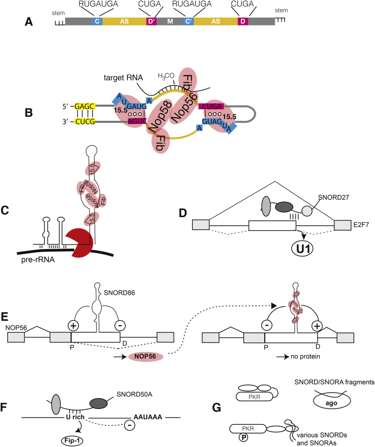
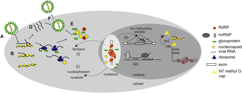
C/D box snoRNAs (SNORDs) are a highly expressed class of non-coding RNAs. Besides their well-established role in rRNA modification, C/D box snoRNAs form protein complexes devoid of fibrillarin and regulate pre-mRNA splicing and polyadenylation of numerous genes. There is an emerging body of evidence for functional interactions between RNA viruses and C/D box snoRNAs. The infectivity of some RNA viruses depends on enzymatically active fibrillarin, and many RNA viral proteins associate with nucleolin or nucleophosmin, suggesting that viruses benefit from their cytosolic accumulation. These interactions are likely reflected by morphological changes in the nucleolus, often leading to relocalization of nucleolar proteins and ncRNAs to the cytosol that are a characteristic feature of viral infections. Knock-down studies have also shown that RNA viruses need specific C/D box snoRNAs for optimal replication, suggesting that RNA viruses benefit from gene expression programs regulated by SNORDs, or that viruses have evolved "new" uses for these humble ncRNAs to advance their prospects during infection.


Figures

Fig. 1
Fig. 1
Molecular functions of snoRNAs. A. Schematic structure of a C/D box snoRNAs. C, D and C′, D′ boxes are indicated with their consensus sequences, AS: antisense boxes, M: middle domain. In most cases SNORDs have short sequences exhibiting complementarity at the ends, which form short stems (see also B). B. Hypothetical structure of human snoRNP performing 2′-O-methylation. The SNORD forms a protein complex made of 15.5 (also known as SNU13 and NHP2L1), NOP56/58 and the methylase fibrillarin (Fib) that 2′-O-methylates (H3CO-) rRNA at a defined position (5 nt upstream of the D box). The coloring of the SNORD is similar to Fig. 1A. Circles indicate the base interaction within the RNA kink. Only one antisense box is shown in rRNA targeting, but both antisense boxes can be used. The structure is adopted from an archea snoRNP, based on NMR and cryo-EM studies [18,19]. C. SNORD3 guides endonucleases (red) to pre-rRNA, leading to cleavage. In addition to the C and D boxes, SNORD3 contains A and B boxes that interact with rRNA. D. Role of SNORDs in pre-mRNA splicing. SNORD27 binds to areas near an alternative 5′ splice site and blocks usage of this site through competition with U1 snRNP [20]. The constitutive exons are in gray, alternative exons are in white, the splicing patterns are indicated. E. Autoregulation of NOP56 formation due to alternative splicing. SNORD86 is located in an alternative 5′ exon (white box) of the NOP56 pre-mRNA. Without NOP56, the SNORD86 structure in the pre-mRNA activates a proximal splice site (P) and represses a distal (D) one, leading to exclusion of the alternative exon and the formation of a mRNA encoding NOP56 (left). The formation of a snoRNP containing NOP56 reverses this regulation, now blocking the proximal splice site and activating the distal one (right). The resulting mRNA does not encode a protein. An intermediate form that still contains R2TP proteins partially represses the proximal splice site (not shown) [21]. F. SNORD50A regulates polyadenylation. SNORD50A binds to the U-rich element that is part of the polyadenylation recognition site. SNORD50 binding removes FIP-1, a component of the cleavage and polyadenylation specificity factor (CPSF), which blocks polyadenylation at this site. Thus removal of SNORD50A increases usage of numerous alternative polyadenylation sites and increases expression of some mRNAs [22]. G. SNORDs bind to proteins. SNORDs and SNORAs bind to protein kinase RNA, leading to its activation, measured by PKR autophosphorylation (P) [23]. Fragments of SNORDs and SNORAs bind to Argonaute proteins, acting as miRNAs.
Fig. 2
Fig. 2
Replication cycle of a generic RNA virus. Although specific steps of viral replication cycle may differ between virus families, the requisite discrete steps are outlined here. A. Virus attaches to the target host cell via interactions of surface exposed glycoproteins (green bars) and host cell surface proteins that serve as viral receptors. B. The virus particle is internalized via endocytosis or the viral genome is released into the cytoplasm via fusion of the viral and cellular membranes. Inside the cell the viral genome, typically coated with a viral nucleocapsid protein (yellow balls), the genomic RNA is transcribed by the viral RNA-dependent RNA polymerase (RDRP; red stars) to make mRNA (for negative sense viruses), or the genome serves directly as mRNA (for positive sense viruses). C. The mRNAs are translated by host cell ribosomes, either in the cytosol, within concentrated areas of viral production (so called virus factories, often associated with the Golgi) or on the rough ER (e.g., for glycoproteins that require further processing). D. The viral genome is replicated with the virally encoded RDRP (RNA dependent RNA polymerase). E. Newly synthesized viral proteins and viral genomes self-assemble into new virus particles. F. New virions are released from the cell via exocytosis, fusion with the cell membrane, or upon cell lysis.Viral components may access host snoRNAs either when the snoRNAs are present in the cytosol, when viral proteins possess nuclear or nucleolar localization signals, or in the special cases when the virus employs a replication strategy that included nuclear localization, e.g., the orthomyxoviruses, bornaviruses, or retroviruses. During infection, viruses can cause components of the SNORD methylating complex to be re-purposed. For example, i. viral infection of some viruses cause translocation of fibrillarin, nucleophosmin or nucleolin from the nucleolus to the cytosol. ii. SNORDs normally employed for rRNA modification in methylation complexes in the nucleolus can be re-trafficked into the nucleoplasm where they influence splice site selection or, iii., polyadenylation and mRNA stability; iv. Nascent transcripts of SNORDs that are independently transcribed can be exploited by viruses for cap-snatching, transferring a N7methyl guanosine cap to the 5′ end of a viral RNA.

Similar articles

Cited by

References

    1. Reddy R., Henning D., Busch H. Nucleotide sequence of nucleolar U3B RNA. J. Biol. Chem. 1979;254:11097–11105. - PubMed
    1. Maxwell E.S., Fournier M.J. The small nucleolar RNAs. Annu. Rev. Biochem. 1995;64:897–934. - PubMed
    1. Tyc K., Steitz J.A. U3, U8 and U13 comprise a new class of mammalian snRNPs localized in the cell nucleolus. EMBO J. 1989;8:3113–3119. - PMC - PubMed
    1. Shapiro E., Biezuner T., Linnarsson S. Single-cell sequencing-based technologies will revolutionize whole-organism science. Nat. Rev. Genet. 2013;14:618–630. - PubMed
    1. Bratkovic T., Rogelj B. The many faces of small nucleolar RNAs. Biochim. Biophys. Acta. 2014;1839:438–443. - PubMed

LinkOut - more resources