

An unusual case of abdominal aneurysm: Inferior vena cava aneurysm - A case report
- PMID: 31193685
- PMCID: PMC6538599
- DOI: 10.1016/j.jccase.2018.08.011
An unusual case of abdominal aneurysm: Inferior vena cava aneurysm - A case report
Abstract
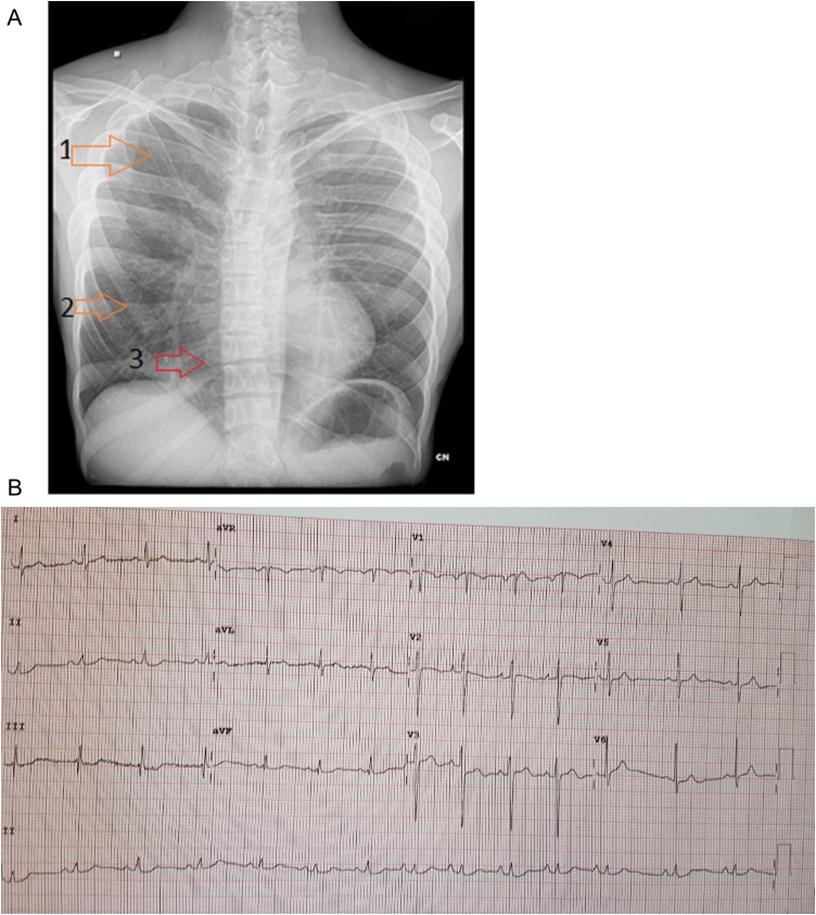
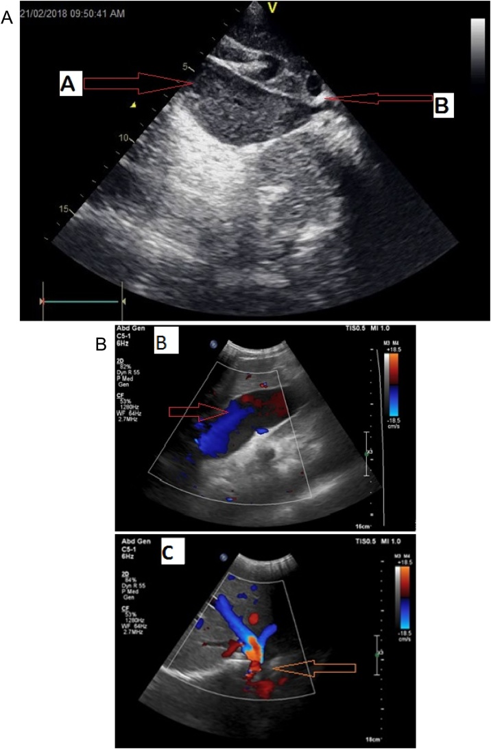
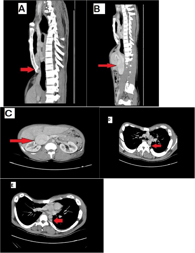
Venous aneurysms are rarely reported in the literature since they are usually asymptomatic and incidentally detected due to complications such as thrombosis and pulmonary embolism. Often an inferior vena cava (IVC) aneurysm is detected by imaging studies performed for other causes. We report a case of large Type II IVC aneurysm associated with severe pectus excavatum in an asymptomatic man detected on routine 2D echocardiography. Focal narrowing of the IVC at the level of xiphisternum detected in multi-slice computed tomography might be the possible etiology for IVC aneurysm. <Learning objective: Even though inferior vena cava aneurysms are rare, they can easily be diagnosed non-invasively by 2D echocardiography, ultrasound, and multi-slice computed tomography. The proximal obstruction of inferior vena cava by the xiphisternum as a complication of a severe form of pectus excavatum resulting in inferior vena cava aneurysm is probably a rare and possibly the only reported case in the literature.>.
Keywords: 2D echocardiography; Aneurysm; Computed tomography; Inferior vena cava.
Figures

References
-
- Oh K.S., Dorst J.P., Haroutunian L.M. Inferior vena caval varix. Radiology. 1973;109:161–162. - PubMed
-
- Montero-Baker M.F., Branco B.C., Leon L.L., Jr., Labropoulos N., Echeverria A., Mills J.L., Sr. Management of inferior vena cava aneurysm. J Cardiovasc Surg (Torino) 2015;56:769–774. - PubMed
-
- Atalar M.H. Aneurysm of the inferior vena cava: imaging findings. Austin J Radiol. 2016;3:1053.
-
- Yalamanchili K., Summer W., Valentine V. Pectus excavatum with inspiratory inferior vena cava compression: a new presentation of pulses paradoxus. Am J Med Sci. 2005;329:45–47. - PubMed
LinkOut - more resources
Full Text Sources
