. 2019 May 21:6:457-464.
doi: 10.1016/j.toxrep.2019.05.008. eCollection 2019.

IL-6 and NFE2L2: A putative role for the hepatoprotective effect of N. Sativa, P. Ginseng and C. Sempervirens in AFB-1 induced hepatocellular carcinoma in rats

Affiliations

IL-6 and NFE2L2: A putative role for the hepatoprotective effect of N. Sativa, P. Ginseng and C. Sempervirens in AFB-1 induced hepatocellular carcinoma in rats

Nora M Aborehab et al. Toxicol Rep. .

Abstract

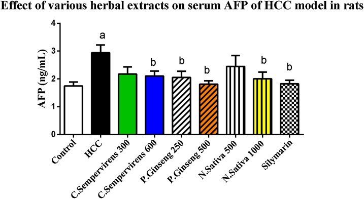
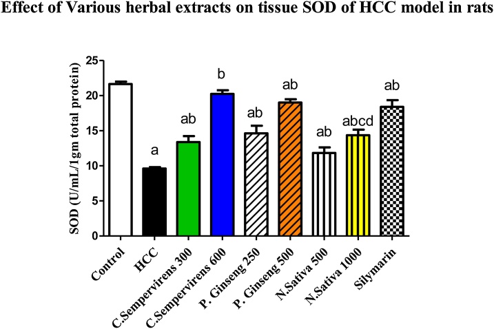
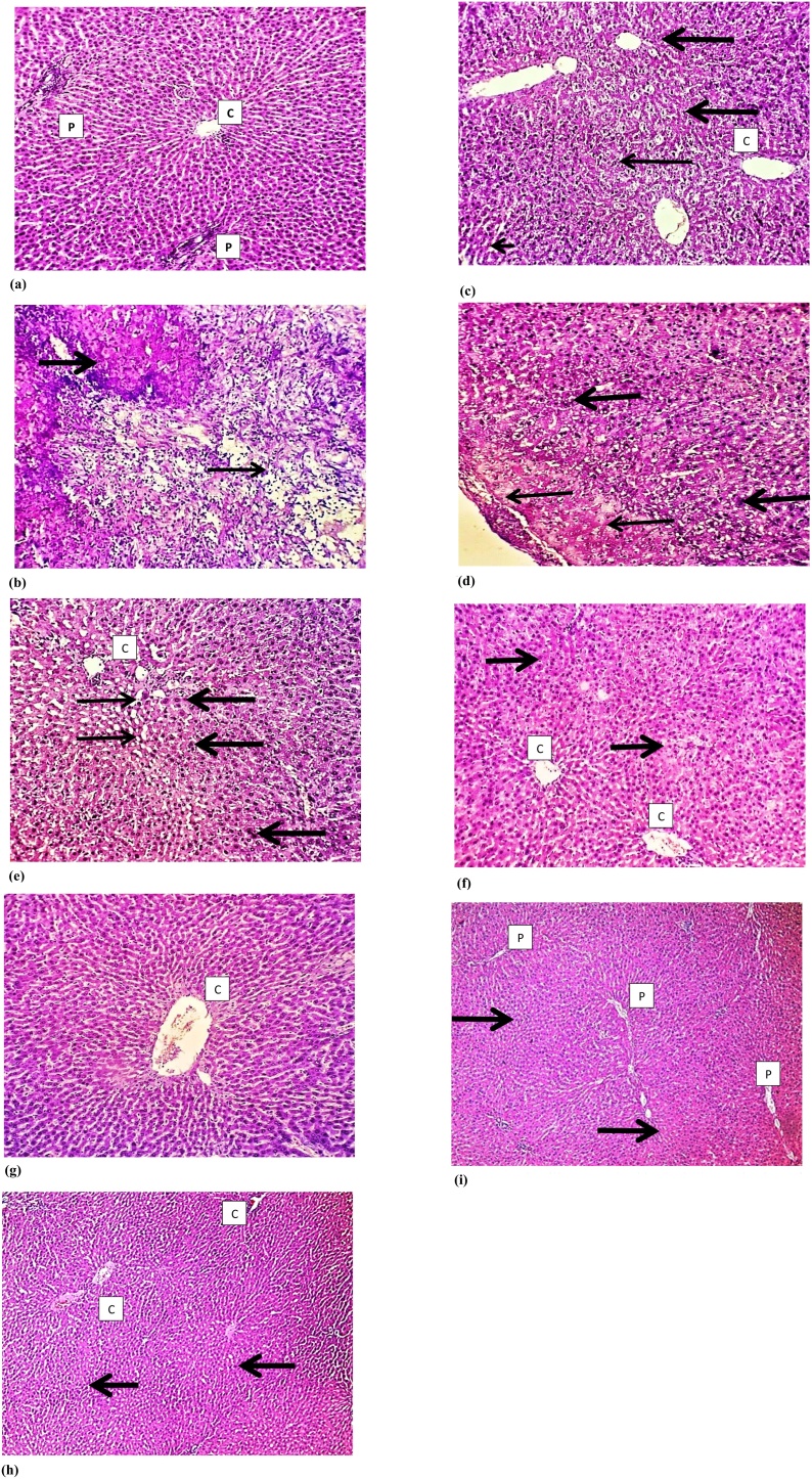
In this study, we investigated possible hepato-protective effects of N. Sativa, P. Ginseng, and C. Sempervirens in Aflatoxin B1 (AFB-1) induced hepatocellular carcinoma rat model. Fifty-four male albino rats were randomly assigned to experimental groups. Alcoholic extracts of aforementioned herbs were administered orally for 28 days at different doses. IL-6, hs-CRP, MDA, SOD and NFE2L2 were determined using ELISA. Histopathological changes in treated groups were examined. Herbal treatment significantly reduced IL-6, hs-CRP, and MDA (P < 0.001) whereas it significantly increased SOD (p < 0.001). C. Sempervirens 600 and N. Sativa 1000 increased NFE2L2 level compared to P. Ginseng 500 group (P value<0.01). Histopathological evaluation of treated groups showed different grades of healing of the liver. This study confirms a beneficial hepatoprotective effect for aforementioned herbal extracts orally administered in rat model of AFB1 induced HCC. This effect is putatively mediated via modulation of inflammatory cytokines as well as amelioration of oxidative stress.

Keywords: C. Sempervirens; HCC; IL-6; N. Sativa; NFE2L2; P. Ginseng.


Figures

None
Graphical abstract
Fig. 1
Fig. 1
Serum level of Alpha-fetoprotein (ng/mL) in the experimental groups. N. Sativa, P. Ginseng and C. Sempervirens extracts reduced serum level of AFP in the liver cancer rats after 1 month of prophylaxis. Results were expressed as mean ± SEM and analyzed using one-way ANOVA followed by Bonferroni’s post hoc test, a: significant from control at P < 0.05, b: significant from AFB1 induced HCC group at P < 0.05
Fig. 2
Fig. 2
Serum level of IL-6 (Pg/mL) in the experimental groups. N. Sativa, P. Ginseng and C. Sempervirens extracts reduced serum level of IL-6 in the liver cancer rats after 1 month of prophylaxis. Results were expressed as mean ± SEM and analyzed using one-way ANOVA followed by Bonferroni’s post hoc test, a: significant from control at P < 0.0001, b: significant from AFB1 induced HCC group at P < 0.0001, c: significant from C. Sempervirens 600 at P < 0.0001, d: significant from P. Ginseng 500 at P < 0.0001
Fig. 3
Fig. 3
Serum level of hs-CRP (ng/mL) in the experimental groups. N. Sativa, P. Ginseng and C. Sempervirens extracts reduced serum level of hs-CRP in the liver cancer rats after 1 month of prophylaxis. Results were expressed as mean ± SEM and analyzed using one-way ANOVA followed by Bonferroni’s post hoc test, a: significant from control at P < 0.01, b: significant from AFB1 induced HCC group at P < 0.001, c: significant from C. Sempervirens 600 at P < 0.05, d: significant from P. Ginseng 500 at P < 0.05.
Fig. 4
Fig. 4
Tissue level of MDA (nmole/mL/1gm total protein) in the experimental groups. N. Sativa, P. Ginseng and C. Sempervirens extracts reduced tissue level of MDA in the liver cancer rats after 1 month of prophylaxis. Results were expressed as mean ± SEM and analyzed using one-way ANOVA followed by Bonferroni’s post hoc test, a: significant from control at P < 0.0001, b: significant from AFB1 induced HCC group at P < 0.0001, c: significant from C. Sempervirens 600 at P < 0.05, d: significant from P. Ginseng 500 at P < 0.05.
Fig. 5
Fig. 5
Tissue level of SOD (U/mL/1 gm total protein) in the experimental groups. N. Sativa, P. Ginseng and C. Sempervirens extracts reduced tissue level of SOD in the liver cancer rats after 1 month of prophylaxis. Results were expressed as mean ± SEM and analyzed using one-way ANOVA followed by Bonferroni’s post hoc test, a: significant from control at P < 0.0001, b: significant from AFB1 induced HCC group at P < 0.0001, c: significant from C. Sempervirens 600 at P < 0.0001, d: significant from P. Ginseng 500 at P < 0.0001
Fig. 6
Fig. 6
Tissue level of NFE2L2 (U/mL/1gm total protein) in the experimental groups. N. Sativa, P. Ginseng and C. Sempervirens extracts raised tissue level of NFE2L2 in the liver cancer rats after 1 month of prophylaxis. Results were expressed as mean ± SEM and analyzed using one-way ANOVA followed by Bonferroni’s post hoc test, a: significant from control at P < 0.0001, b: significant from AFB1 induced HCC group at P < 0.0001, d: significant from P. Ginseng 500 at P < 0.01
Fig. 7
Fig. 7
Histological examination of the liver tissue of all experimental groups using Hematoxylin and Eosin (H&E) staining: a) illustrates control group sections that shows normal hepatocytes and preserved hepatic lobular architecture, with regular central veins (C) and unremarkable portal tracts (P). b) Illustrates Aflatoxin group section that shows wide neoplastic transformation of the hepatocytes, with increased N/C ratio, nuclear hyperchromasia and irregular nuclear membranes. c) Illustrates Silymarin treated group section that shows preservation of the lobular architecture, subserosal hepatocyte dysplastic changes is focally evident, however with no frank neoplastic transformation. d) Illustrates N. Sativa group sections that shows evident preservation of the lobular architecture, minimal hepatocyte dysplastic changes is focally evident (thick arrow), however with no neoplastic transformation. e) and f) illustrates C. Sempervirens and P. Ginseng group sections that shows evident preservation of the lobular architecture with unremarkable hepatocyte dysplastic changes and no neoplastic transformation. Magnification 10 × .

Similar articles

Cited by

References

    1. Ahmed Mohammed H.A. Factors Influencing Surveillance for Hepatocellular Carcinoma in Patients with Liver Cirrhosis. Liver Cancer. 2017;6(2):126–136. - PMC - PubMed
    1. Siegel R.L., Miller K.D., Jemal A. Cancer statistics, 2016. CA Cancer J Clin. 2016;66(1):7–30. - PubMed
    1. Liu Z. Synergisic effect of APRIL knockdown and Jiedu Xiaozheng Yin, a Chinese medicinal recipe, on the inhibition of hepatocellular carcinoma cell proliferation. Oncol Rep. 2017;37(2):754–760. - PMC - PubMed
    1. Anwar W.A. Changing pattern of hepatocellular carcinoma (HCC) and its risk factors in Egypt: possibilities for prevention. Mutat Res. 2008;659(1-2):176–184. - PubMed
    1. Abdel-Hamid N.M. Can methanolic extract of Nigella sativa seed affect glyco-regulatory enzymes in experimental hepatocellular carcinoma? Environ Health Prev Med. 2013;18(1):49–56. - PMC - PubMed