

Fitz-Hugh-Curtis syndrome resulting in nutmeg liver on computed tomography
- PMID: 31193761
- PMCID: PMC6542378
- DOI: 10.1016/j.radcr.2019.04.008
Fitz-Hugh-Curtis syndrome resulting in nutmeg liver on computed tomography
Abstract
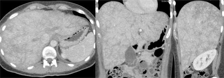
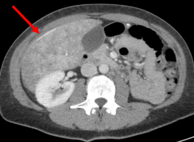
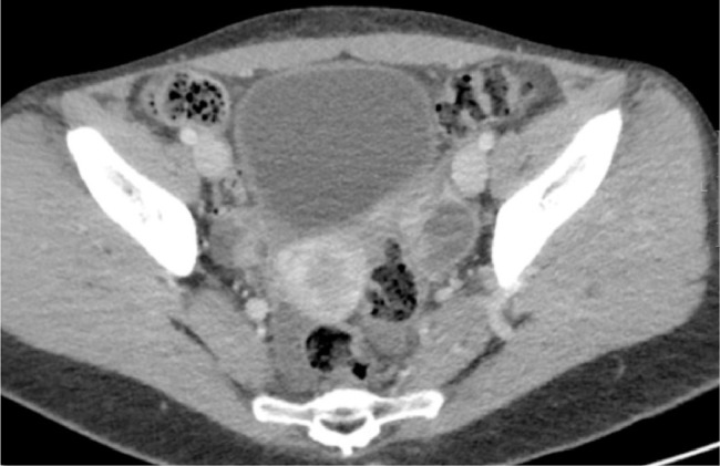
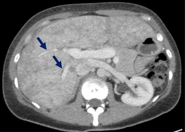
A 34-year-old woman entered the emergency room with abdominal pain in the right upper quadrant. Computed tomography scan showed a nutmeg liver suspected for increased venous pressure by thrombosis of the liver veins, Budd-Chiari malformation, or right-sided heart failure. Interestingly, the diagnosis was pelvic inflammatory disease complicated by the Fitz-Hugh-Curtis syndrome (FHCS). Pelvic inflammatory disease resulted from an ascended infection by Chlamydia trachomatis. FHCS was caused by perihepatitis defined as inflammation of the peritoneal capsule of the liver. Fast diagnosis and treatment is crucial. Therefore, we report a case of FHCS characterized by a nutmeg liver on computed tomography.
Keywords: Chlamydia trachomatis; FHCS, Fitz-Hugh-Curtis syndrome; Fitz-Hugh-Curtis syndrome; GP, general practitioner; Hepatomegaly; IUD, intrauterine device; Nutmeg liver; PID, pelvic inflammatory disease; Pelvic Inflammatory disease; Perihepatitis.
Figures
Similar articles
-
Fitz-Hugh-Curtis syndrome: A cause of right upper quadrant abdominal pain.Med Clin (Barc). 2020 Jun 12;154(11):447-452. doi: 10.1016/j.medcli.2020.01.022. Epub 2020 Mar 4. Med Clin (Barc). 2020. PMID: 32145988 Review. English, Spanish.
-
Fitz-Hugh-Curtis syndrome in a man positive for Chlamydia trachomatis.Clin J Gastroenterol. 2018 Aug;11(4):338-342. doi: 10.1007/s12328-018-0829-5. Epub 2018 Feb 7. Clin J Gastroenterol. 2018. PMID: 29417387 Review.
-
Right pleural effusion in Fitz-Hugh-Curtis syndrome.Acta Med Okayama. 2006 Oct;60(5):289-94. doi: 10.18926/AMO/30742. Acta Med Okayama. 2006. PMID: 17072375
-
Fitz Hugh Curtis Case Report.J Educ Teach Emerg Med. 2020 Apr 15;5(2):V19-V21. doi: 10.21980/J82K9G. eCollection 2020 Apr. J Educ Teach Emerg Med. 2020. PMID: 37465405 Free PMC article.
-
A rare case of Fitz-Hugh-Curtis syndrome caused by Chlamydia trachomatis in an HIV-positive male patient.SAGE Open Med Case Rep. 2019 Jan 16;7:2050313X18823592. doi: 10.1177/2050313X18823592. eCollection 2019. SAGE Open Med Case Rep. 2019. PMID: 30728975 Free PMC article.
Cited by
-
Fitz-Hugh-Curtis syndrome: a case of perihepatitis in 'mosaic' pattern.BMJ Case Rep. 2022 Mar 2;15(3):e248744. doi: 10.1136/bcr-2022-248744. BMJ Case Rep. 2022. PMID: 35236709 Free PMC article. No abstract available.
References
-
- Mishori R., McClaskey E.L., WinklerPrins V.J. Chlamydia trachomatis infections: screening, diagnosis, and management. Am Fam Physician. 2012;86(12):1127–1132. - PubMed
-
- Wiesenfeld H.C., Hillier S.L., Krohn M.A., Amortegui A.J., Heine R.P., Landers D.V. Lower genital tract infection and endometritis: insight into subclinical pelvic inflammatory disease. Obstet Gynecol. 2002;100(3):456–463. - PubMed
-
- Kobayashi Y., Takeuchi H., Kitade M., Kikuchi I., Sato Y., Kinoshita K. Pathological study of Fitz-Hugh-Curtis syndrome evaluated from fallopian tube damage. J Obstet Gynaecol Res. 2006;32(3):280–285. - PubMed
-
- Paavonen J., Eggert-Kruse W. Chlamydia trachomatis: impact on human reproduction. Hum Reprod Update. 1999;5(5):433–447. - PubMed
-
- Peter N.G., Clark L.R., Jaeger J.R. Fitz-Hugh-Curtis syndrome: a diagnosis to consider in women with right upper quadrant pain. Cleve Clin J Med. 2004;71:233–239. - PubMed
Publication types
LinkOut - more resources
Full Text Sources
