

An inverted appendix found on routine colonoscopy: A case report with discussion of imaging findings
- PMID: 31193843
- PMCID: PMC6543180
- DOI: 10.1016/j.radcr.2019.05.022
An inverted appendix found on routine colonoscopy: A case report with discussion of imaging findings
Abstract
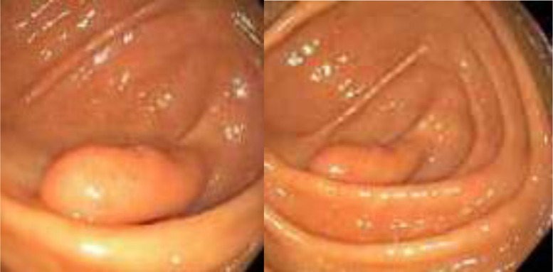
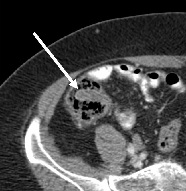
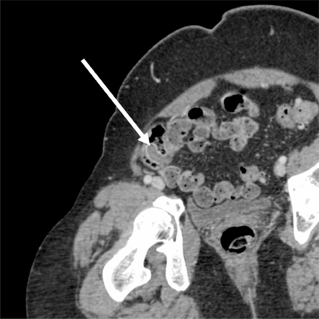
Appendiceal inversion is an uncommon incidental finding on colonoscopy that can mimic pathologic processes such as colon polyps and neoplasms due to its mass-like appearance. Endoscopic removal of these lesions has been associated with a higher risk of peritonitis and bleeding. Awareness of appendiceal inversion may potentially decrease unwarranted interventions as well as its associated risks. Although there are many reported cases of iatrogenic appendiceal inversion due to the traditional inversion-ligation technique performed during open appendectomy, there are few reported cases of asymptomatic appendiceal inversion without a known history of iatrogenic inversion. Here, we present a case of an asymptomatic patient with appendiceal inversion and no prior history of appendectomy. Furthermore, we discuss management and characteristic imaging findings of appendiceal inversion that may help to distinguish it from similarly appearing pathologic conditions.
Keywords: Appendiceal inversion; Colonoscopy; Inverted appendix.
Figures
References
-
- Järvensivu P., Lehtola J., Karvonen A.L., Rinne A., Suramo I. Colonoscopic appearance of the remnant of the appendix after total inversion. Endoscopy. 1982;14:66–68. - PubMed
- Järvensivu P., Lehtola J., Karvonen A.L., Rinne A., Suramo I.. Colonoscopic appearance of the remnant of the appendix after total inversion. Endoscopy. 1982;14. 66–8. 10.1055/s-2007-1021581. - PubMed
-
- Engström L., Fenyö G. Appendicectomy: assessment of stump invagination versus simple ligation: a prospective, randomized trial. Br J Surg. 1985;72:971–972. - PubMed
- Engström L., Fenyö G.. Appendicectomy: assessment of stump invagination versus simple ligation: a prospective, randomized trial. Br J Surg, 1985;72: 971–2. doi:10.1002/bjs.1800721212 - PubMed
Publication types
LinkOut - more resources
Full Text Sources
