

Imbalanced development of anterior and posterior thorax is a causative factor triggering scoliosis
- PMID: 31194037
- PMCID: PMC6551366
- DOI: 10.1016/j.jot.2018.12.001
Imbalanced development of anterior and posterior thorax is a causative factor triggering scoliosis
Abstract
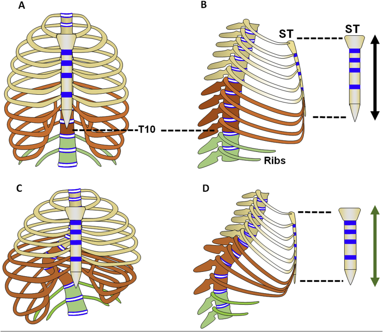
Objective: Scoliosis is a common disease characterized by spinal curvature with variable severities. There is no generally accepted theory about the physical origin of the spinal deformation of scoliosis. The aim of this study was to explore a new hypothesis suggesting that the curvatures in scoliosis may be associated with the imbalance growth between thoracic vertebral column and sternum.
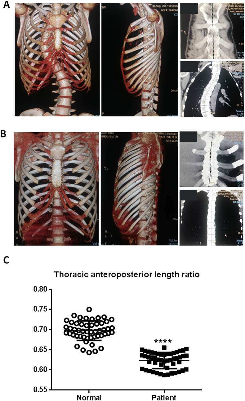
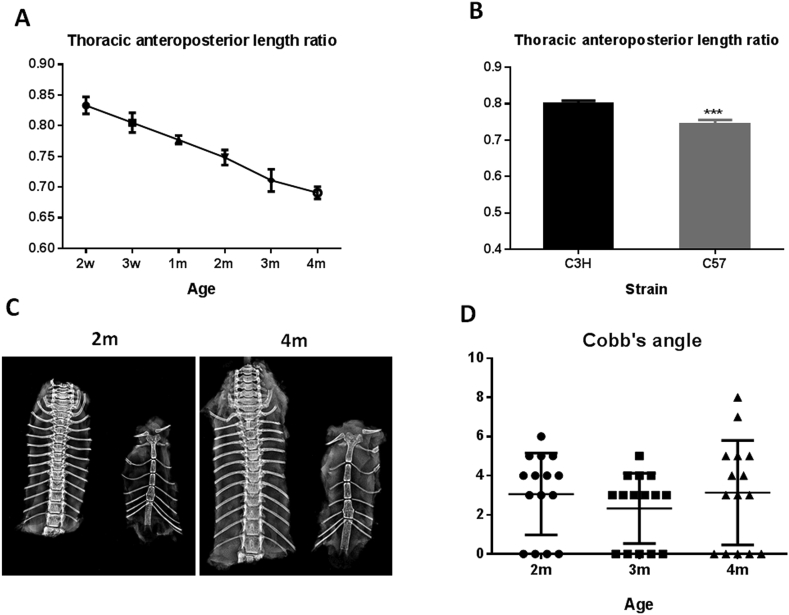
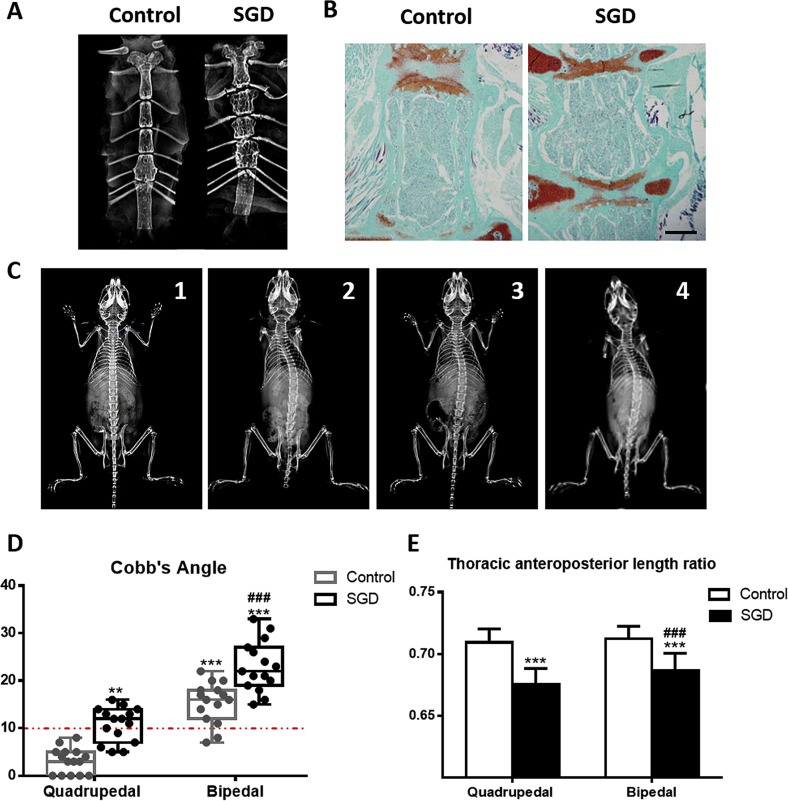
Methods: We undertook a comparative computed tomography (CT) based morphology study of thoracic vertebrae and sternum of patients with adolescent idiopathic scoliosis (AIS) and age-gender matched normal subjects. We further measured the ratios between the lengths of the sternum and thoracic vertebra of mice with deficiency of fibroblast growth factor receptor 3 (FGFR3), which exhibit scoliosis. Three-week-old C57BL/6J mice were used to generate bipedal and sternal growth plate injury model. Radiographs and histological images were obtained to observe the presence of sternal and spinal deformity.
Results: There was a significant correlation between the severities of scoliosis and the ratios of the sternum to thoracic vertebral lengths. We also found that FGFR3 deficient mice showed smaller ratio of the sternum to thoracic vertebra lengths than that of the wild-type mice, which were similar with that of the AIS patients. Surgery-induced injuries of sternal growth plates can accelerate and aggravate the scoliosis in bipedal mice and imbalanced development of anterior and posterior thoracic occurred before the appearance of scoliosis.
Conclusions: Our findings suggest that the imbalanced growth between the thoracic vertebral column and the sternum is an important causative factor for the pathogenesis of scoliosis including AIS.
The translational potential of this article: Imbalanced growth between the thoracic vertebral column and the sternum is associated with scoliosis. Surgical or rehabilitation intervention for scoliosis should focus on all components involved in the pathogenesis of curvature to obtain better outcome.
Keywords: Growth plate; Imbalanced growth; Scoliosis; Sternum; Vertebrae.
Figures

Similar articles
-
Anterior-posterior length discrepancy of the spinal column in adolescent idiopathic scoliosis-a 3D CT study.Spine J. 2018 Dec;18(12):2259-2265. doi: 10.1016/j.spinee.2018.05.005. Epub 2018 May 4. Spine J. 2018. PMID: 29730457
-
The posterior skeletal thorax: rib-vertebral angle and axial vertebral rotation asymmetries in adolescent idiopathic scoliosis.Stud Health Technol Inform. 2008;140:263-8. Stud Health Technol Inform. 2008. PMID: 18810034
-
The effectiveness of selective thoracic fusion for treating adolescent idiopathic scoliosis: a systematic review protocol.JBI Database System Rev Implement Rep. 2015 Nov;13(11):4-16. doi: 10.11124/jbisrir-2015-2338. JBI Database System Rev Implement Rep. 2015. PMID: 26657460
-
Anterior Spinal Overgrowth of the Thoracic Spine May Not Be Involved in the Initiation of Adolescent Idiopathic Scoliosis.World Neurosurg. 2019 May;125:e319-e325. doi: 10.1016/j.wneu.2019.01.071. Epub 2019 Jan 25. World Neurosurg. 2019. PMID: 30685373
-
Restrained Differential Growth: The Initiating Event of Adolescent Idiopathic Scoliosis?Spine (Phila Pa 1976). 2017 Jun 15;42(12):E726-E732. doi: 10.1097/BRS.0000000000001946. Spine (Phila Pa 1976). 2017. PMID: 27792114 Review.
Cited by
-
Incidentally Detected Pericardial Defect in a Patient with Pneumothorax as Confirmed on Video-Assisted Thoracoscopic Surgery.Taehan Yongsang Uihakhoe Chi. 2021 May;82(3):749-755. doi: 10.3348/jksr.2020.0057. Epub 2021 Mar 22. Taehan Yongsang Uihakhoe Chi. 2021. PMID: 36238774 Free PMC article.
-
Patients with Adolescent Idiopathic Scoliosis Have Higher Metabolic Cost during High-Intensity Interval Training.Int J Environ Res Public Health. 2023 Jan 25;20(3):2155. doi: 10.3390/ijerph20032155. Int J Environ Res Public Health. 2023. PMID: 36767522 Free PMC article.
-
Letter to the editor concerning "Imbalanced development of anterior and posterior thorax is a causative factor triggering scoliosis" by Chen et al., Journal of Orthopaedic Translation, 2019, https://doi.org/10.1016/j.jot.2018.12.001.J Orthop Translat. 2019 Aug 27;22:142. doi: 10.1016/j.jot.2019.07.008. eCollection 2020 May. J Orthop Translat. 2019. PMID: 32440510 Free PMC article. No abstract available.
-
In vivo kinematic study of lumbar center of rotation under different loads.BMC Musculoskelet Disord. 2025 Feb 14;26(1):155. doi: 10.1186/s12891-025-08410-8. BMC Musculoskelet Disord. 2025. PMID: 39953502 Free PMC article.
-
Electromyographic Analysis of Paraspinal Muscles of Scoliosis Patients Using Machine Learning Approaches.Int J Environ Res Public Health. 2022 Jan 21;19(3):1177. doi: 10.3390/ijerph19031177. Int J Environ Res Public Health. 2022. PMID: 35162203 Free PMC article.
References
-
- Altaf F., Gibson A., Dannawi Z., Noordeen H. Adolescent idiopathic scoliosis. BMJ. 2013;346:f2508. - PubMed
-
- Burton D.C., Carlson B.B., Place H.M., Fuller J.E., Blanke K., Cho R. Results of the scoliosis Research society morbidity and mortality database 2009–2012: a report from the morbidity and mortality committee. Spine Deformity. 2016;4(5):338–343. - PubMed
-
- Cho W., Shepard N., Arlet V. The etiology of congenital scoliosis: genetic vs. environmental-a report of three monozygotic twin cases. Eur Spine J. 2018;27(Suppl 3):533–537. - PubMed
-
- Weinstein S.L., Dolan L.A., Cheng J.C., Danielsson A., Morcuende J.A. Adolescent idiopathic scoliosis. Lancet. 2008;371(9623):1527–1537. - PubMed
LinkOut - more resources
Full Text Sources
Research Materials
