

Bioactive scaffolds for osteochondral regeneration
- PMID: 31194079
- PMCID: PMC6551354
- DOI: 10.1016/j.jot.2018.11.006
Bioactive scaffolds for osteochondral regeneration
Abstract
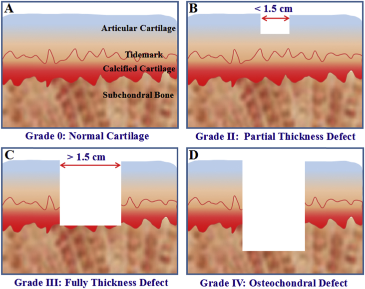
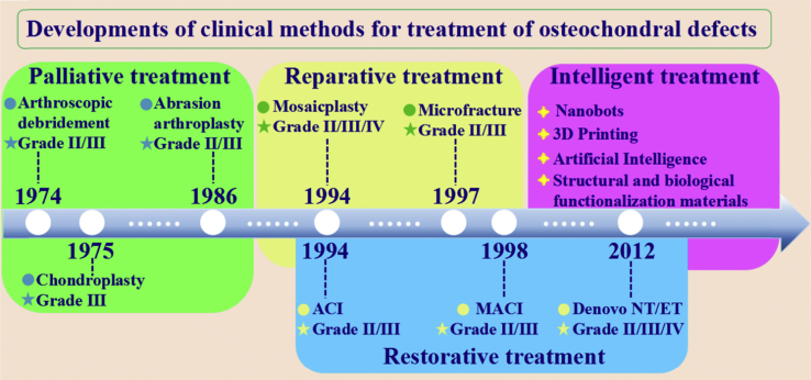
Treatment for osteochondral defects remains a great challenge. Although several clinical strategies have been developed for management of osteochondral defects, the reconstruction of both cartilage and subchondral bone has proved to be difficult due to their different physiological structures and functions. Considering the restriction of cartilage to self-healing and the different biological properties in osteochondral tissue, new therapy strategies are essential to be developed. This review will focus on the latest developments of bioactive scaffolds, which facilitate the osteogenic and chondrogenic differentiation for the regeneration of bone and cartilage. Besides, the topic will also review the basic anatomy, strategies and challenges for osteochondral reconstruction, the selection of cells, biochemical factors and bioactive materials, as well as the design and preparation of bioactive scaffolds. Specifically, we summarize the most recent developments of single-type bioactive scaffolds for simultaneously regenerating cartilage and subchondral bone. Moreover, the future outlook of bioactive scaffolds in osteochondral tissue engineering will be discussed. This review offers a comprehensive summary of the most recent trend in osteochondral defect reconstruction, paving the way for the bioactive scaffolds in clinical therapy.
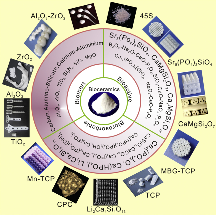
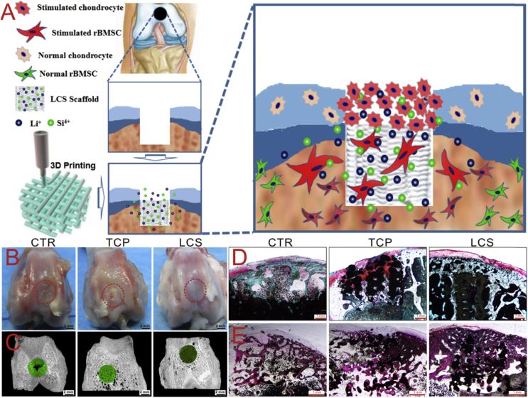
The translational potential of this article: This review summaries the latest developments of single-type bioactive scaffolds for regeneration of osteochondral defects. We also highlight a new possible translational direction for cartilage formation by harnessing bioactive ions and propose novel paradigms for subchondral bone regeneration in application of bioceramic scaffolds.
Keywords: Bioactive scaffolds; Bioceramics; Cartilage repair; Osteochondral regeneration; Subchondral bone regeneration.
Figures
Similar articles
-
Advances of nanotechnology in osteochondral regeneration.Wiley Interdiscip Rev Nanomed Nanobiotechnol. 2019 Nov;11(6):e1576. doi: 10.1002/wnan.1576. Epub 2019 Jul 22. Wiley Interdiscip Rev Nanomed Nanobiotechnol. 2019. PMID: 31329375 Review.
-
Bioactive Scaffolds for Regeneration of Cartilage and Subchondral Bone Interface.Theranostics. 2018 Feb 15;8(7):1940-1955. doi: 10.7150/thno.23674. eCollection 2018. Theranostics. 2018. PMID: 29556366 Free PMC article.
-
Osteochondral Tissue Engineering Dilemma: Scaffolding Trends in Regenerative Medicine.Stem Cell Rev Rep. 2023 Aug;19(6):1615-1634. doi: 10.1007/s12015-023-10545-x. Epub 2023 Apr 19. Stem Cell Rev Rep. 2023. PMID: 37074547 Review.
-
Treatment of osteochondral defects in the rabbit's knee joint by implantation of allogeneic mesenchymal stem cells in fibrin clots.J Vis Exp. 2013 May 21;(75):e4423. doi: 10.3791/4423. J Vis Exp. 2013. PMID: 23728213 Free PMC article.
-
Cryogenic 3D printing of heterogeneous scaffolds with gradient mechanical strengths and spatial delivery of osteogenic peptide/TGF-β1 for osteochondral tissue regeneration.Biofabrication. 2020 Mar 23;12(2):025030. doi: 10.1088/1758-5090/ab7ab5. Biofabrication. 2020. PMID: 32106097
Cited by
-
The role of the immune microenvironment in bone, cartilage, and soft tissue regeneration: from mechanism to therapeutic opportunity.Mil Med Res. 2022 Nov 19;9(1):65. doi: 10.1186/s40779-022-00426-8. Mil Med Res. 2022. PMID: 36401295 Free PMC article. Review.
-
3D bioprinted scaffolds for osteochondral regeneration: advancements and applications.Mater Today Bio. 2025 May 8;32:101834. doi: 10.1016/j.mtbio.2025.101834. eCollection 2025 Jun. Mater Today Bio. 2025. PMID: 40487176 Free PMC article. Review.
-
3D printed osteochondral scaffolds: design strategies, present applications and future perspectives.Front Bioeng Biotechnol. 2024 Feb 15;12:1339916. doi: 10.3389/fbioe.2024.1339916. eCollection 2024. Front Bioeng Biotechnol. 2024. PMID: 38425994 Free PMC article. Review.
-
Osteochondral Injury, Management and Tissue Engineering Approaches.Front Cell Dev Biol. 2020 Nov 4;8:580868. doi: 10.3389/fcell.2020.580868. eCollection 2020. Front Cell Dev Biol. 2020. PMID: 33251212 Free PMC article. Review.
-
Evaluation of Glycerylphytate Crosslinked Semi- and Interpenetrated Polymer Membranes of Hyaluronic Acid and Chitosan for Tissue Engineering.Polymers (Basel). 2020 Nov 11;12(11):2661. doi: 10.3390/polym12112661. Polymers (Basel). 2020. PMID: 33187239 Free PMC article.
References
-
- Murphy L., Helmick C.G. The impact of osteoarthritis in the United States: a population-health perspective: a population-based review of the fourth most common cause of hospitalization in U.S. adults. Orthop Nurs. 2012;112(1):85–91. - PubMed
-
- Cheng Y.J., Hootman J.M., Murphy L.B., Langmaid G.A., Helmich C.G. Prevalence of doctor-diagnosed arthritis and arthritis-attributable activity limitation - United States, 2007-2009. Mmwr Morb Mortal Wkly Rep. 2010;59(39):1261–1265. - PubMed
-
- Re’Em T., Witte F., Willbold E., Ruvinov E., Cohen S. Simultaneous regeneration of articular cartilage and subchondral bone induced by spatially presented TGF-beta and BMP-4 in a bilayer affinity binding system. Acta Biomater. 2012;8(9):3283–3293. - PubMed
Publication types
LinkOut - more resources
Full Text Sources
Other Literature Sources
Research Materials
