

MRI myelography for diagnosis and targeted blood patching of multilevel thoracic spine CSF leaks: Report of 2 cases
- PMID: 31194096
- PMCID: PMC6551536
- DOI: 10.1016/j.radcr.2019.05.006
MRI myelography for diagnosis and targeted blood patching of multilevel thoracic spine CSF leaks: Report of 2 cases
Abstract
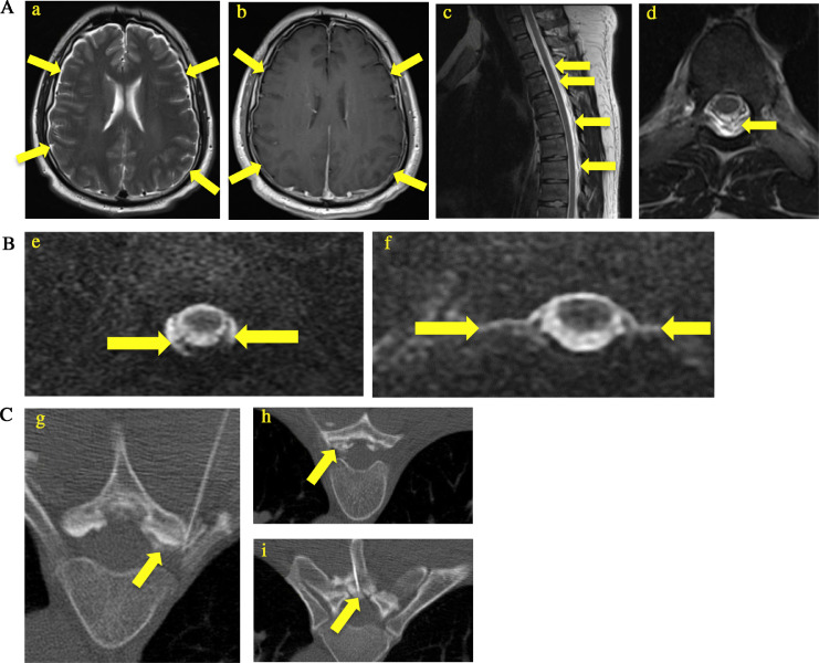
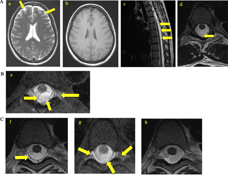
In patients with occult cerebrospinal fluid (CSF) leaks or CSF leak syndrome, orthostatic headaches are a common presenting symptom. Although computed tomography (CT) myelography has historically been the gold standard for diagnosis with radioisotope cisternography as a diagnostic alternative, magnetic resonance imaging (MRI) myelography using intrathecal gadolinium has reported sensitivity of 80%-87%. Two patients with spontaneous orthostatic headaches lasting for several days were diagnosed with CSF leaks at multiple thoracic segments using MRI myelogram with intrathecal gadolinium (Gadavist, Bayer, Whippany, NJ). This allowed for subsequent targeted treatment with CT fluoroscopy guidance, resulting in therapeutic responses within 1-2 treatment with targeted blood patching. Although intrathecal gadolinium is an off-label use, the superior contrast resolution and lack of radiation exposure makes MRI myelography an excellent imaging modality for diagnosing CSF leak, targeting treatment, and monitoring outcomes compared to CT myelography and radioisotope cisternography.
Keywords: Blood patch; CSF leak; MR myelogram.
Figures
Similar articles
-
MR myelography for identification of spinal CSF leak in spontaneous intracranial hypotension.AJNR Am J Neuroradiol. 2014 Oct;35(10):2007-12. doi: 10.3174/ajnr.A3975. Epub 2014 May 22. AJNR Am J Neuroradiol. 2014. PMID: 24852289 Free PMC article.
-
The utility of radioisotope cisternography in low CSF/volume syndromes compared to myelography.Cephalalgia. 2016 Nov;36(13):1291-1295. doi: 10.1177/0333102416628467. Epub 2016 Sep 30. Cephalalgia. 2016. PMID: 26823556 Clinical Trial.
-
Leakage detection on CT myelography for targeted epidural blood patch in spontaneous cerebrospinal fluid leaks: calcified or ossified spinal lesions ventral to the thecal sac.J Neurosurg Spine. 2014 Sep;21(3):432-41. doi: 10.3171/2014.5.SPINE13549. Epub 2014 Jun 20. J Neurosurg Spine. 2014. PMID: 24949904
-
Spontaneous Intracranial Hypotension: Imaging in Diagnosis and Treatment.Radiol Clin North Am. 2019 Mar;57(2):439-451. doi: 10.1016/j.rcl.2018.10.004. Epub 2018 Dec 7. Radiol Clin North Am. 2019. PMID: 30709479 Review.
-
Spontaneous CSF leaks: low CSF volume syndromes.Neurol Clin. 2014 May;32(2):397-422. doi: 10.1016/j.ncl.2013.11.002. Epub 2014 Feb 28. Neurol Clin. 2014. PMID: 24703536 Review.
Cited by
-
T2-Sampling Perfection With Application-Optimized Contrasts by Using Flip Angle Evolution (SPACE) Protocol MRI: A Safe, Minimally Invasive Screening Tool for Spinal CSF Leak Causing Spontaneous Intracranial Hypotension.Cureus. 2022 Jul 7;14(7):e26626. doi: 10.7759/cureus.26626. eCollection 2022 Jul. Cureus. 2022. PMID: 35949747 Free PMC article.
References
-
- Kranz P.G., Luetmer P.H., Diehn F.E., Amrhein T.J., Tanpitukpongse T.P., Gray L. Myelographic techniques for the detection of spinal CSF leaks in spontaneous intracranial hypotension. Am J Roentgenol. 2016;206(1):8–19. - PubMed
- Kranz P.G., Luetmer P.H., Diehn F.E., Amrhein T.J., Tanpitukpongse T.P., Gray L.Myelographic techniques for the detection of spinal CSF leaks in spontaneous intracranial hypotension. Am J Roentgenol. 2016;206(1):8–19. doi:10.2214/AJR.15.14884 - PubMed
-
- Algin O., Turkbey B. Intrathecal gadolinium-enhanced MR cisternography: a comprehensive review. Am J Neuroradiol. 2013;34(1):14–22. - PMC - PubMed
- Algin O., Turkbey B.Intrathecal gadolinium-enhanced MR cisternography: A a comprehensive review. Am J Neuroradiol. 2013;34(1):14–22. doi:10.3174/ajnr.A2899 - PMC - PubMed
-
- Chazen J, Talbott J, Lantos J, Dillon W. MR myelography for identification of spinal CSF leak in spontaneous intracranial hypotension. Am J Neuroradiol. 2014;35(10):2007–2012. - PMC - PubMed
- Chazen J, Talbott J, Lantos J, Dillon W. MR myelography for identification of spinal CSF leak in spontaneous intracranial hypotension. Am J Neuroradiol. 2014;35(10):2007–12. - PMC - PubMed
-
- Selcuk H., Albayram S., Ozer H. Intrathecal gadolinium-enhanced MR cisternography in the evaluation of CSF leakage. Am J Neuroradiol. 2010;31(1):71–75. - PMC - PubMed
- Selcuk H., Albayram S., Ozer H., et al. Intrathecal gadolinium-enhanced MR cisternography in the evaluation of CSF leakage. Am J Neuroradiol. 2010;31(1):71–5. doi:10.3174/ajnr.A1788 - PMC - PubMed
Publication types
LinkOut - more resources
Full Text Sources
