. 2019 Dec 2;20(23):2927-2935.
doi: 10.1002/cbic.201900276. Epub 2019 Sep 20.

Modulation and Visualization of EF-G Power Stroke During Ribosomal Translocation

Affiliations

Modulation and Visualization of EF-G Power Stroke During Ribosomal Translocation

Heng Yin et al. Chembiochem. .

Abstract

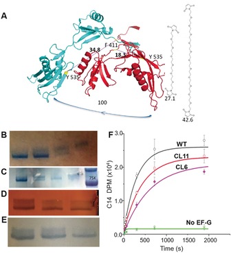
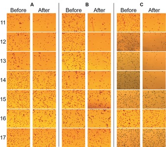
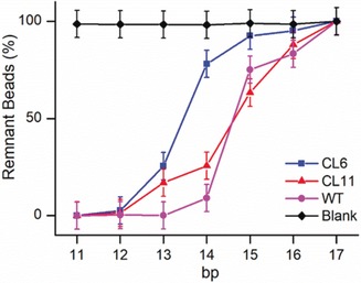
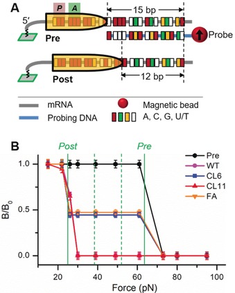
During ribosome translocation, the elongation factor EF-G undergoes large conformational change while maintaining its contact with the moving tRNA. We previously measured a power stroke accompanying EF-G catalysis, which was consistent with structural studies. However, the role of power stroke in translocation fidelity remains unclear. Here, we report quantitative measurements of the power strokes of structurally modified EF-Gs by using two different techniques and reveal the correlation between power stroke and translocation efficiency and fidelity. We discovered that the reduced power stroke only lowered the percentage of translocation but did not introduce translocation error. The established force -structure-function correlation for EF-G indicates that power stroke drives ribosomal translocation, but the mRNA reading frame is probably maintained by ribosome itself. Furthermore, the microscope detection method reported here can be simply implemented for other biochemical applications.

Keywords: antibiotics; crosslinked EF-G; force-induced remnant magnetization spectroscopy; power stroke; ribosomal translocation; translocation fidelity.


Conflict of interest statement

The authors declare no conflict of interest.

Figures

Figure 1
Figure 1
Design, purification, and activity assay of crosslinked EF‐Gs. A) Crosslinking residues F411 and Y535 (bacterial numbering). B) Separation by continuous elution electrophoresis. Lanes 1 and 4: pure lower and upper bands, respectively; lanes 2 and 3: the transitions from one component to the other. Lanes are numbered from left to right. C) Purification by MiniGel and hand‐incision, followed by Bio‐Rad Model 422 elution. Lanes 1 and 3: purified CL EF‐G with different quantities; lane 2: mixture of un‐crosslinked and crosslinked; lane 4: purified un‐crosslinked EF‐G; lane 5: marker. D) Purification by activated thiol Sepharose 4B. Lanes 1–3 were analyzed after 1 h, 4 h, and overnight incubation time with the beads, respectively. The final purity was approximately 70 %. E) Purification by maleimide‐activated magnetic beads. Lanes 1–3 were analyzed after 1, 2, and 3 h of incubation time, respectively. The final purity was >90 %. F) Radioactivity measurements by the poly(Phe) assay for the WT and two crosslinked EF‐Gs.
Figure 2
Figure 2
Magnetic method for measuring the power stroke of crosslinked EF‐Gs. A) Schematic of using DNA–mRNA duplexes as force rulers and magnetic labeling to determine the power stroke. The equal and opposite forces are reminiscent to myosin–actin interaction. B) Plot of remnant magnetic beads versus duplex length for CL11. C) Plot of remnant magnetic beads versus duplex length for CL6. D) Overlay of the EF‐G structures before and after translocation to indicate the extraordinary conformational changes. Red: domain IV; blue: the rest domains.
Figure 3
Figure 3
Microscope images for determining the power stroke of crosslinked and wild‐type EF‐Gs. A) CL6, B) CL11, C) WT EF‐G, 11–17: base pair number of the DNA–mRNA rulers.
Figure 4
Figure 4
Results of particle counting for the microscope detection of the EF‐G power stroke. Blank was the control with no EF‐G.
Figure 5
Figure 5
Reduced power stroke of EF‐G bound with fusidic acid. A) Plot of remnant magnetic beads versus duplex base pairs. WT EF‐G without fusidic acid is also shown for comparison. FA: fusidic acid. B, C) Representative microscope images after the power stroke for the 12 (B) and 15 bp rulers (C).
Figure 6
Figure 6
Translocation efficiency probed by FIRMS. A) Probing scheme for translocation of the same ribosome complex as in the power stroke experiments. B) Translocation products for different EF‐Gs. The two solid lines indicate the positions of Post and Pre, respectively. The two dashed green lines indicate the expected positions of “−1” (left) and “−2” (right) frameshifting products. Both were absent in the results.

References

    1. Dunkle J. A., Cate J. H. D., Annu. Rev. Biophys. 2010, 39, 227–244. - PubMed
    1. Katunin V. I., Savelsbergh A., Rodnina M. V., Wintermeyer W., Biochemistry 2002, 41, 12806–12812. - PubMed
    1. Achenbach J., Nierhaus K. H., Biochimie 2015, 114, 80–89. - PubMed
    1. Zhou J., Lancaster L., Donohue J. P., Noller H. F., Science 2014, 345, 1188–1191. - PMC - PubMed
    1. Lin J., Gagnon M. G., Bulkley D., Steitz T. A., Cell 2015, 160, 219–227. - PMC - PubMed

Publication types

LinkOut - more resources