. 2019 Jul 11;62(13):6262-6275.
doi: 10.1021/acs.jmedchem.9b00566. Epub 2019 Jun 27.

PeIA-5466: A Novel Peptide Antagonist Containing Non-natural Amino Acids That Selectively Targets α3β2 Nicotinic Acetylcholine Receptors

Affiliations

PeIA-5466: A Novel Peptide Antagonist Containing Non-natural Amino Acids That Selectively Targets α3β2 Nicotinic Acetylcholine Receptors

Arik J Hone et al. J Med Chem. .

Abstract

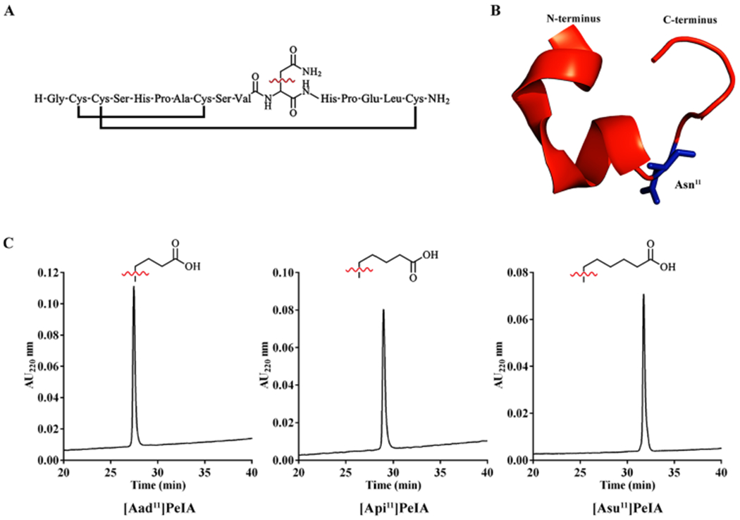
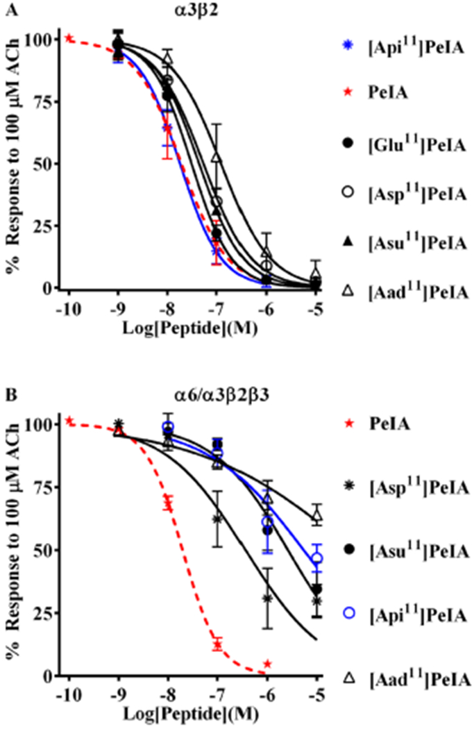
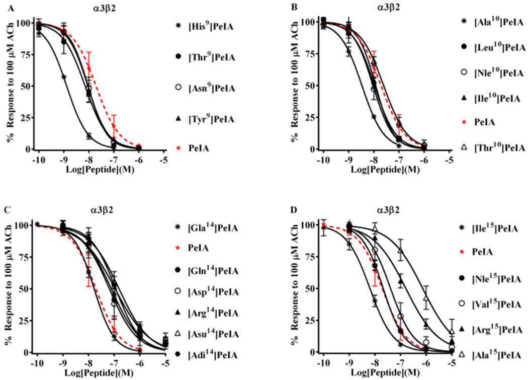
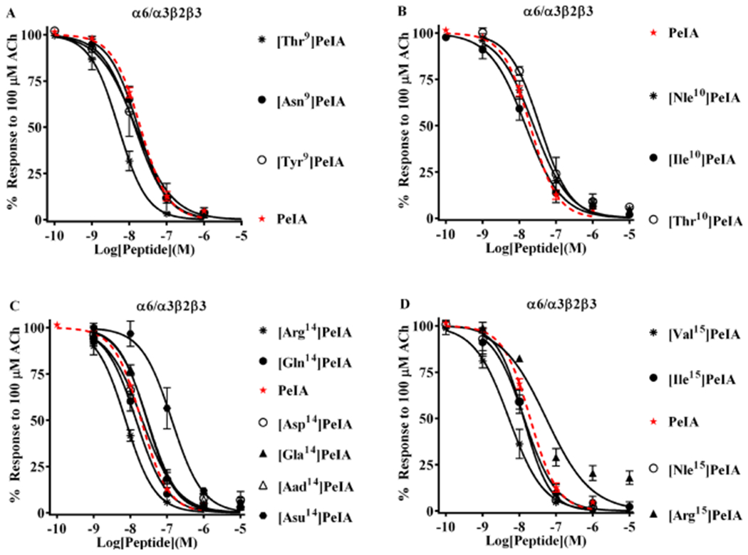
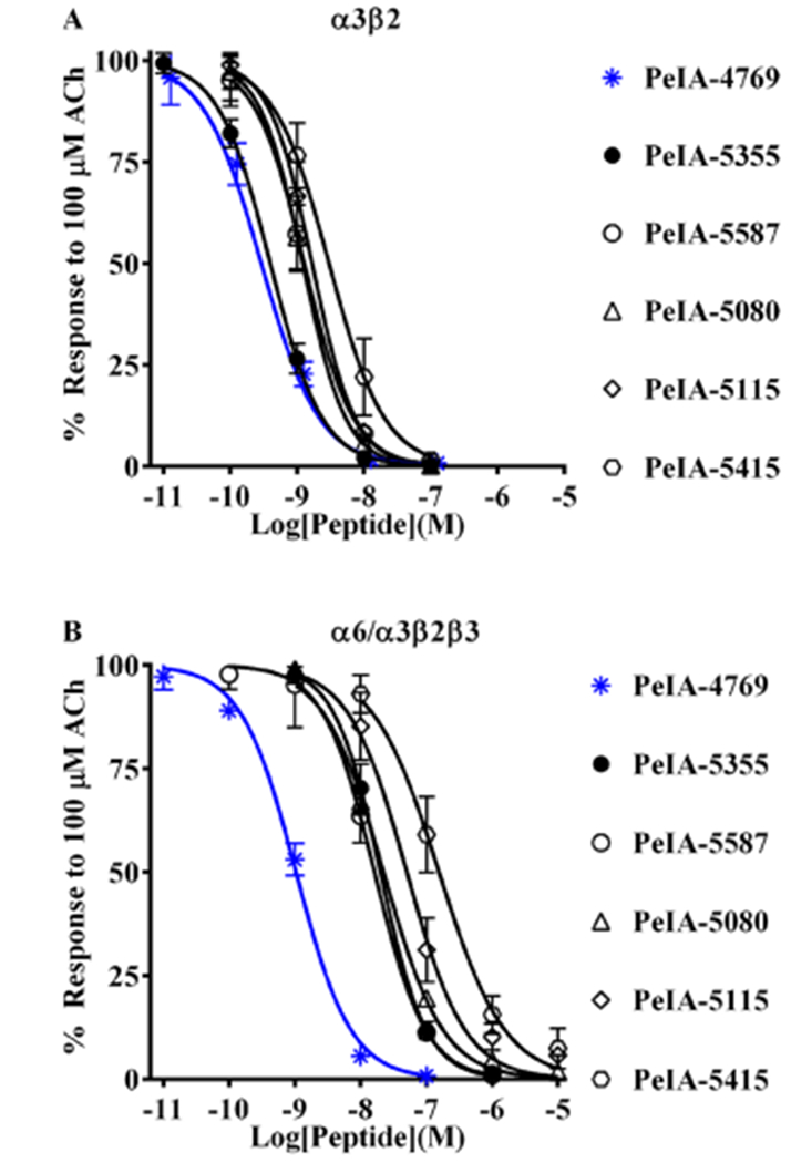
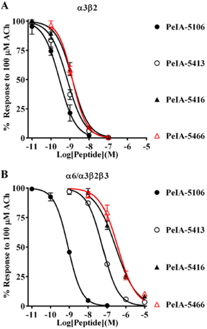
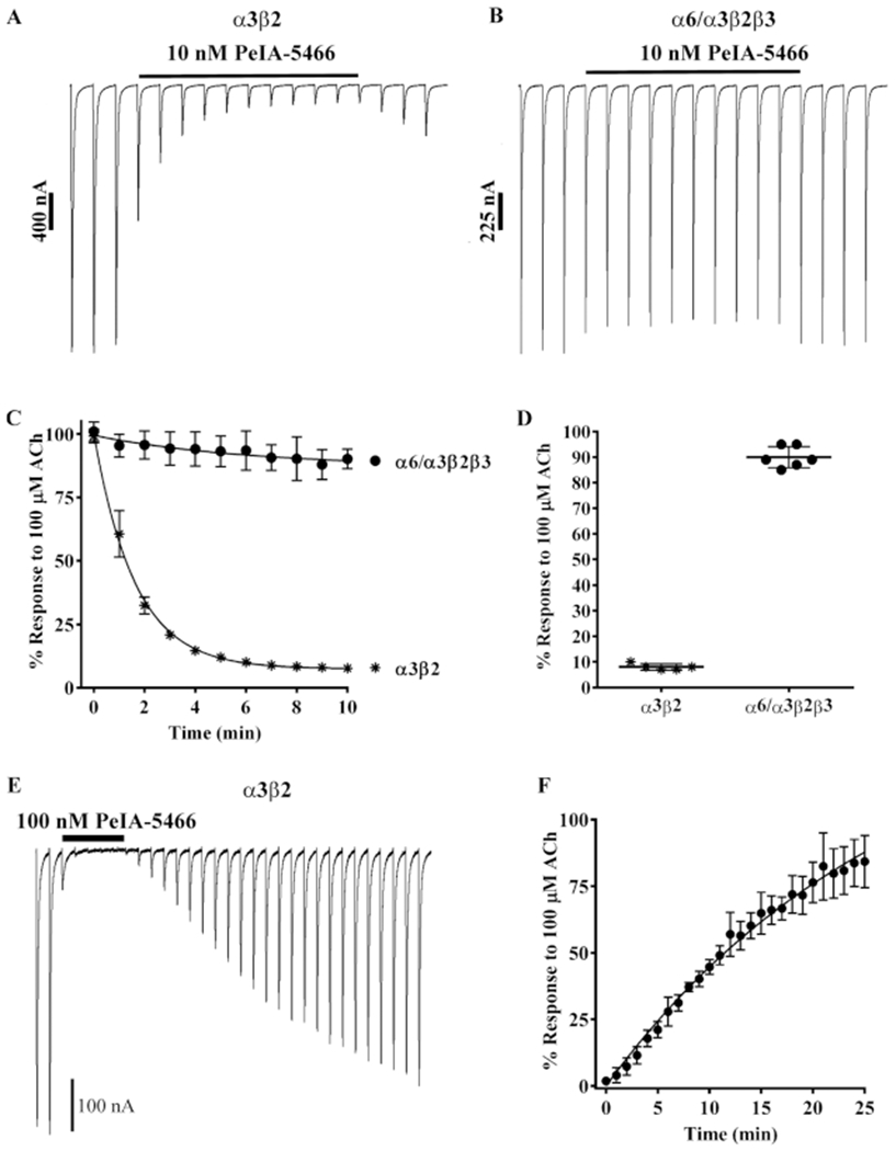
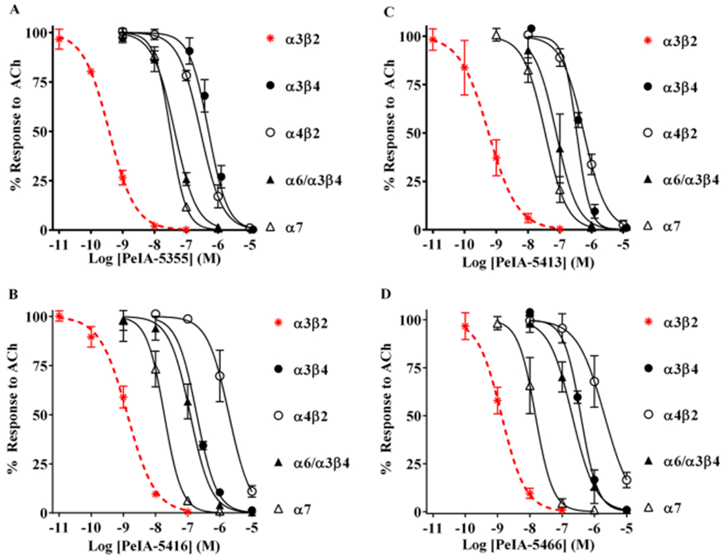
Pharmacologically distinguishing α3β2 nicotinic acetylcholine receptors (nAChRs) from closely related subtypes, particularly α6β2, has been challenging due to the lack of subtype-selective ligands. We created analogs of α-conotoxin (α-Ctx) PeIA to identify ligand-receptor interactions that could be exploited to selectively increase potency and selectivity for α3β2 nAChRs. A series of PeIA analogs were synthesized by replacing amino acid residues in the second disulfide loop with standard or nonstandard residues and assessing their activity on α3β2 and α6/α3β2β3 nAChRs heterologously expressed in Xenopus laevis oocytes. Asparagine11 was found to occupy a pivotal position, and when replaced with negatively charged amino acids, selectivity for α3β2 over α6/α3β2β3 nAChRs was substantially increased. Second generation peptides were then designed to further improve both potency and selectivity. One peptide, PeIA-5466, was ∼300-fold more potent on α3β2 than α6/α3β2β3 and is the most α3β2-selective antagonist heretofore reported.


Conflict of interest statement

The authors declare no competing financial interest

Figures

Figure 1.
Figure 1.
Amino acid sequence and structure of PeIA and RP-HPLC of analogs containing non-natural amino acid residues. (A) The linear sequence of PeIA shown with the disulfide connectivity and Asn11 as the line structure. (B) Cartoon rendition of the crystal structure (PDB: 5JME, chain H) of PeIA highlighting the position of Asn11 (blue). (C) Side-chain structures of the non-natural amino acids Aad, Api, and Asu used in analog synthesis and corresponding RP-HPLC traces of the fully folded peptides. [Aad11]PeIA, [Api11]PeIA, and [Asu11]PeIA eluted at 37.5, 39.0, and 42.4% solvent B, respectively, using an ACN gradient of 10–50% in 40 min and a C18 analytical column.
Figure 2.
Figure 2.
PeIA analogs with negatively charged amino acid residues in position 11 show increased selectivity for α3β2 nAChRs. Analogs with substitutions of Asn11 were synthesized and a concentration–response analysis was performed for each to determine the IC50 values. The value obtained for each analog was compared to that of the native peptide (red dashed curves) to assess the effects of the substitution. (A) Analogs with Asp, Glu, or Api substitutions showed <3-fold change in potency for α3β2 nAChRs. Aad, with a one-carbon longer side chain than Glu and one-carbon shorter than Api, reduced PeIA potency by 6-fold. (B) By contrast, all analogs showed substantially reduced potency (6- to 274-fold) for α6/α3β2β3 nAChRs. The most α3β-selective of these analogs was determined to be [Api11]PeIA (blue curve), which showed a 300-fold preference for the α3β2 subtype. A minimum of four oocytes was used for each IC50 determination, and the error bars indicate the SD. The analogs are listed in rank order of potency from top (most potent) to bottom (least potent); values are presented in Table 3.
Figure 3.
Figure 3.
Structure–function analysis of PeIA identifies amino acid substitutions that increase potency for α3β2 nAChRs. PeIA analogs with substitutions of Ser9, Val10, Glu14, or Leu15 were synthesized and a concentration–response analysis was performed for each to determine the IC50 values. The value for each analog was compared to that of the native peptide to evaluate the interaction between the substituted positions and α3β2 nAChRs. (A) Analogs with Asn, Tyr, or Thr substitutions of Ser9 showed a <3-fold change in potency for α3β2 nAChRs. Substitution of Ser9 with His increased the potency of PeIA by 15-fold. (B) Substitution of Val10 with Ala increased (6-fold) PeIA potency, whereas other hydrophobic amino acids showed a <3-fold change but trended toward increased potency. Substitution with Thr also showed a <3-fold change. (C) Substitutions of Glu14 revealed the importance of a Glu in this position for PeIA potency on α3β2 nAChRs. Substitution with any other negatively charged amino acid reduced potency by 3- to 10-fold. A <3-fold change in potency was observed by changing Glu to Gla. Substitution of Glu14 with Gln or Arg reduced PeIA potency by 4- and 6-fold, respectively. (D) Varying effects on PeIA potency were observed when Leu15 was substituted with other hydrophobic amino acids, whereas Ile increased PeIA potency by 3-fold, Ala reduced potency by 38-fold; Nle and Val produced a <3-fold change. Arg reduced PeIA potency by 8-fold. A minimum of four oocytes was used for each IC50 determination and the error bars indicate the SD. The analogs are listed in rank order of potency from top (most potent) to bottom (least potent); values are provided in Table 4. Data for PeIA (red dashed curves) were previously presented in Figure 2 and shown for the ease of comparison to the curves of the analogs.
Figure 4.
Figure 4.
Structure–function analysis of PeIA identifies amino acid substitutions that decrease potency for α6/α3β2β3 nAChRs. A concentration-response analysis was performed for the newly synthesized PeIA analogs, and the IC50 values compared to that of the native peptide and the IC50 values for α3β2 nAChRs. (A) Analogs with substitutions of Ser9 displayed changes in potencies similar to those for α3β2 nAChRs. [Thr9]PeIA displayed increased (4-fold) potency for α6/α3β2β3 nAChRs, whereas all others showed <3-fold change. Note that none of the Ser9 substitutions changed α3β2-α6/α3β2β3 selectivity. (B) Very little change in potency was observed for the newly synthesized analogs with substitutions of Val10. (C) In contrast to the results obtained for α3β2 nAChRs, α6/α3β2β3 nAChRs were less sensitive to perturbations of the interaction between PeIA position 14 and the receptor. Of the analogs with other negatively charged amino acids, only [Asu14]PeIA showed a change in potency (7-fold loss). Substitution of Glu14 with Gln showed a very little effect (<3-fold) on PeIA potency for α6/α3β2β3 nAChRs, whereas Arg reduced the IC50 value by 3-fold. (D) Like α3β2, varying effects on PeIA potency were also observed for α6/α3β2β3 nAChRs when Leu15 was substituted with other hydrophobic amino acids. Whereas the L15I substitutions increased the IC50 value for α3β2, a <3-fold change was observed for α6/α3β2β3 nAChRs. The L15Nle substitution resulted in a <3-fold change in the IC50, whereas L15V decreased the IC50 for α6/α3β2β3 nAChRs by 4-fold. A minimum of four oocytes was used for each IC50 determination and the error bars indicate the SD. The analogs are listed in rank order of potency from top (most potent) to bottom (least potent); values are provided in Table 4. Data for PeIA (red dashed curves) were previously presented in Figure 2 and shown for the ease of comparison to the curves of the analogs.
Figure 5.
Figure 5.
Novel peptides containing the non-natural amino acids Aad, Api, or Asu show enhanced selectivity for α3β2 over α6/α3β2β3 nAChRs. Based on the structure–activity results with PeIA, a series of peptides were synthesized and a concentration–response analysis was performed for each to determine the IC50 values for the inhibition of α3β2 and α6/α3β2β3 nAChRs as well as the selectivity ratio. (A) The first peptide synthesized, PeIA-4769, shares many of the residues found in PeIA (see Table 5) but has His and Leu residues in positions 9 and 10, respectively. PeIA-4769 was 68-fold more potent (blue curve) than PeIA on α3β2 nAChRs. Five additional peptides, based on PeIA-4769, were synthesized containing non-natural amino acid residues in position 11 and of them PeIA-5355, with Api in position 11, was the most potent (pM IC50 value) on α3β2 nAChRs. (B) The IC50 values for the peptides in (A) were determined for the inhibition of α6/α3β2β3 nAChRs and the α3β2-α6/α3β2β3 selectivity ratios calculated. PeIA-4769 was also potent (nM IC50 value, blue curve) α6/α3β2β3 nAChRs. PeIA-5355 displayed the highest selectivity ratio (59-fold) for α3β2 and α6/α3β2β3 nAChRs. Selectivity ratios of 52- and 39-fold were also observed with PeIA-5415 and PeIA-5587, respectively. A minimum of four oocytes was used for each IC50 determination and the error bars indicate the SD; values are provided in Table 5.
Figure 6.
Figure 6.
PeIA-5466 is potent and highly selective for α3β2 over α6/α3β2β3 nAChRs. Peptides based on PeIA-4769 and PeIA-5355 were synthesized with different residues in the 10th and 15th positions and a concentration–response analysis was performed for each. The IC50 values for inhibition of α3β2 and α6/α3β2β3 nAChRs for each peptide were compared to those of PeIA and the α3β2-α6/α3β2β3 selectivity ratio calculated. (A, B) PeIA-5106 and PeIA-5416 were synthesized to evaluate the effects of Nle10 and Ile15, respectively, on α3β2-α6/α3β2β3 nAChR selectivity. PeIA-5106 was 69-fold more potent than PeIA on α3β2 nAChRs, but only a minor increase (3fold) in α3β2-α6/α3β2β3 selectivity, over PeIA-4769, was found. PeIA-5416 with Ile15, was 220-fold selective for α3β2 nAChRs. An additional 70-fold enhancement of selectivity was obtained with PeIA-5466 (red curves). A minimum of 4 oocytes was used for each IC50 determination and the error bars indicate the SD; values are provided in Table 6.
Figure 7.
Figure 7.
Kinetic analysis of PeIA-5466 for α3β2 and α6/α3β2β3 nAChRs. Xenopus oocytes expressing α3β2 or α6/α3β2β3 nAChRs were subjected to TEVC electrophysiology as described in Methods. (A) Representative current traces from an oocytes expressing α3β2 nAChRs before, during, and after exposure to 10 nM PeIA-5466. (B) Representative current traces from an oocyte expressing α6/α3β2β3 nAChRs before, during, and after exposure to 10 nM PeIA-5466. (C) The exponential fit of the data for the inhibition of α3β2 nAChRs yielded a kobs of 0.617 ± 0.023 min−1 and a t1/2 of 1.1 (1.0–1.2) min. (C, D) The current amplitudes in the presence of 10 nM PeIA-5466 were inhibited to 8 ± 1% (n = 5, SD) of control values for α3β2 nAChRs and to 90 ± 4% (n = 6, SD) of control values for α6/α3β2β3 nAChRs. (E) Representative current traces from an oocyte expressing α3β2 nAChRs showing the inhibition and recovery kinetics during and after exposure to 100 nM PeIA-5466. The current amplitudes in the presence of 100 nM peptide were inhibited to 2 ± 1% (n = 4, SD) of control values. (F) The exponential fit of the data for recovery from inhibition yielded an observed off-rate constant (koff) of 0.065 ± 0.001 min−1 and a t1/2 for recovery of 10.6 (10.2–11.0) min. The Kd was determined to be 1.18 ± 0.47 nM. The 30 s traces in A, B, and E are shown concatenated omitting the 30 s intersweep interval for brevity. The error bars in (C, D, and F) indicate the SD. The Kd was calculated from Kd = koff/kon where kon = (kobskoff)/[ligand].
Figure 8.
Figure 8.
Selectivity profiles of peptides containing non-natural amino acid residues. (A-D) The IC50 values of PeIA-5106, PeIA-5413, PeIA-5416, and PeIA-5466 were determined for a panel of nAChR subtypes including α3β4, α4β2, α6/α3β4, and α7. PeIA-5355 displayed the largest selectivity ratio for α3β2 and α3β4 subtypes (1329-fold). PeIA-5466 displayed the largest selectivity ratio between α3β2 nAChRs and both α4β2 and α6/α3β4 subtypes. A minimum of four oocytes was used for each IC50 curve with the exception of those for α3β4 where three oocytes were used. The exceptionally slow on-rate kinetics of all peptides for α3β4 nAChRs required 45–60 min to reach equilibrium using a concentration of 1 μM (data not shown). Two successive applications of 10 μM were then applied immediately after the equilibrium was achieved with 1 μM. The long perfusion time required for concentrations <300 nM exceeded practical recording times and only a few data points were obtained (<3). The error bars indicate the SD; values are provided in Table 6. Data for α3β2 nAChRs (dashed red curves) were previously presented in Figure 6 and shown for the ease of comparison to the curves for other subtypes.

Similar articles

Cited by

References

    1. Gotti C; Zoli M; Clementi F Brain nicotinic acetylcholine receptors: native subtypes and their relevance. Trends Pharmacol. Sci 2006, 27, 482–491. - PubMed
    1. Hone AJ; McIntosh JM Nicotinic acetylcholine receptors in neuropathic and inflammatory pain. FEBS Lett. 2018, 592, 1045–1062. - PMC - PubMed
    1. Brunzell DH Preclinical evidence that activation of mesolimbic alpha6 subunit containing nicotinic acetylcholine receptors supports nicotine addiction phenotype. Nicotine Tob. Res 2012, 14, 1258–1269. - PMC - PubMed
    1. Alexander SP; Peters JA; Kelly E; Marrion N; Benson HE; Faccenda E; Pawson AJ; Sharman JL; Southan C; Davies JA CGTP Collaborators. The concise guide to PHARMACOLOGY 2015/16: ligand-gated ion channels. Br. J. Pharmacol 2015, 172, 5870–5903. - PMC - PubMed
    1. Baddick CG; Marks MJ An autoradiographic survey of mouse brain nicotinic acetylcholine receptors defined by null mutants. Biochem. Pharmacol 2011, 82, 828–841. - PMC - PubMed

Publication types