Review
. 2019 Dec 18;31(1):1-57.
doi: 10.1515/revneuro-2018-0096.

Brain structure and cognitive ability in healthy aging: a review on longitudinal correlated change

Affiliations
Review

Brain structure and cognitive ability in healthy aging: a review on longitudinal correlated change

Jessica Oschwald et al. Rev Neurosci. .

Abstract

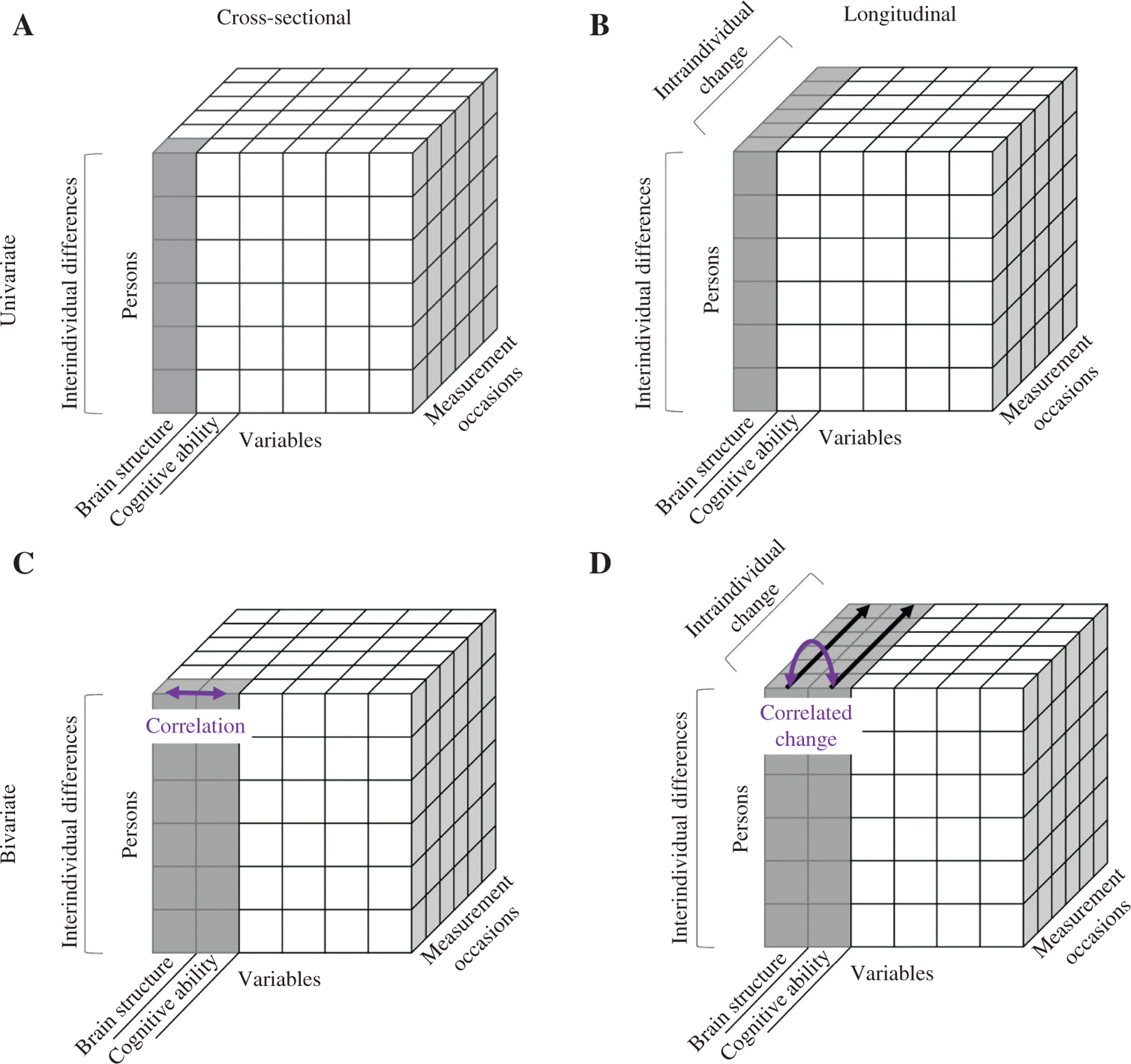
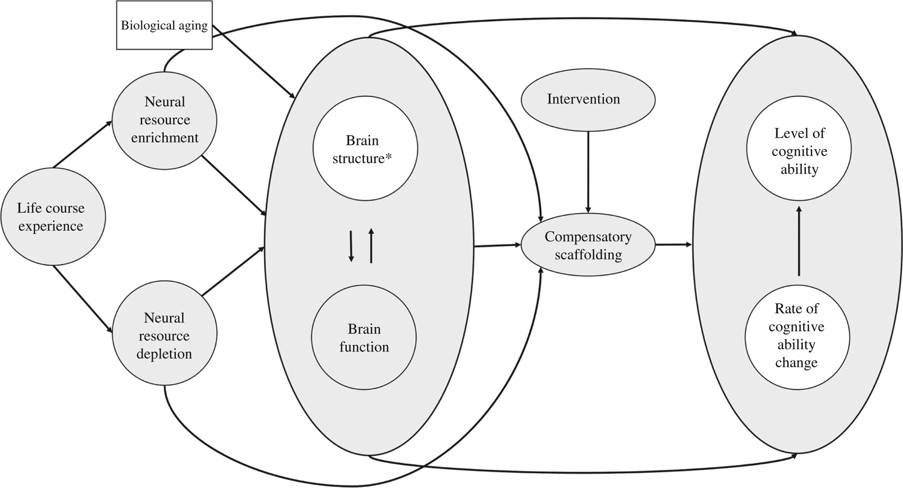
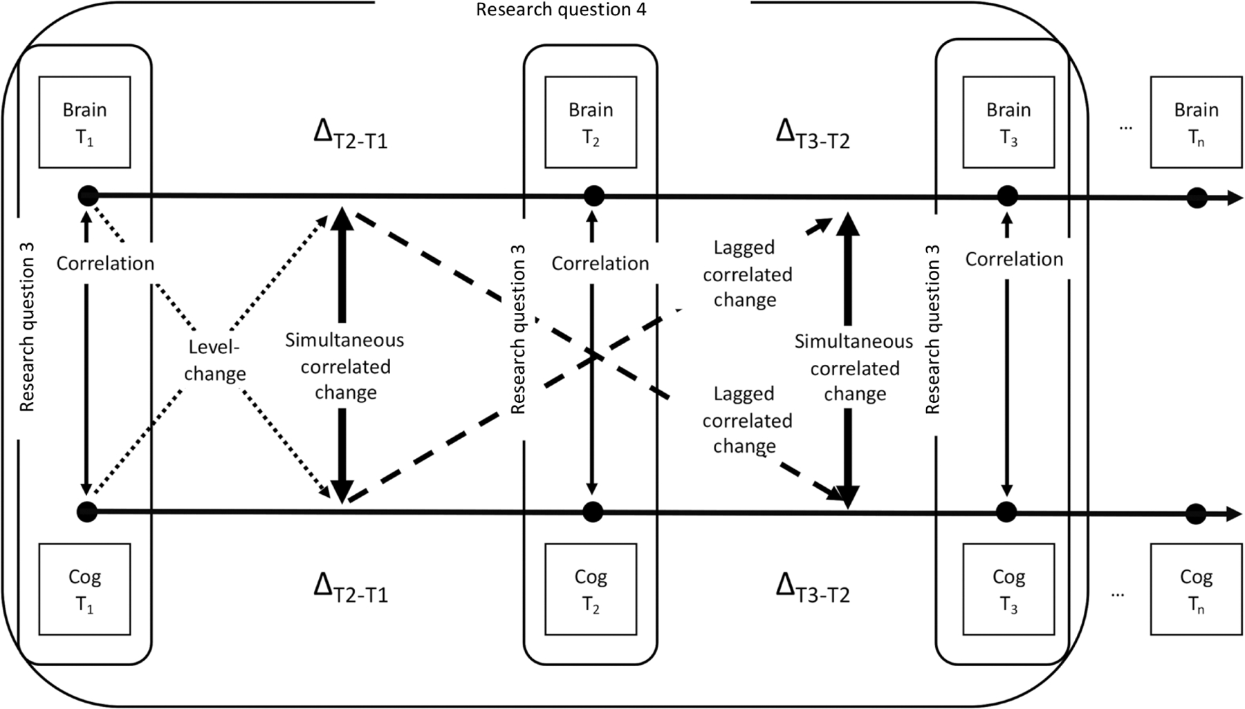
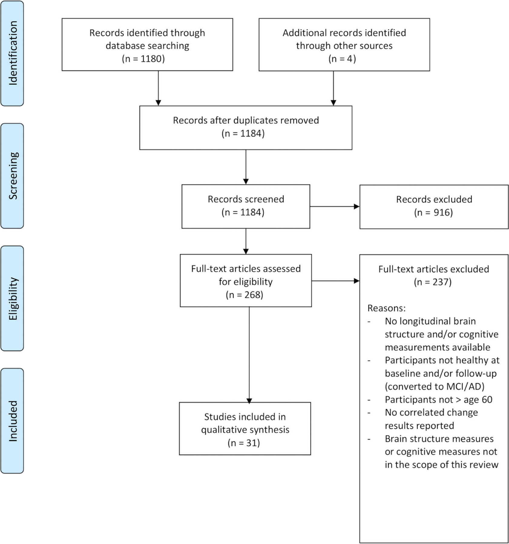
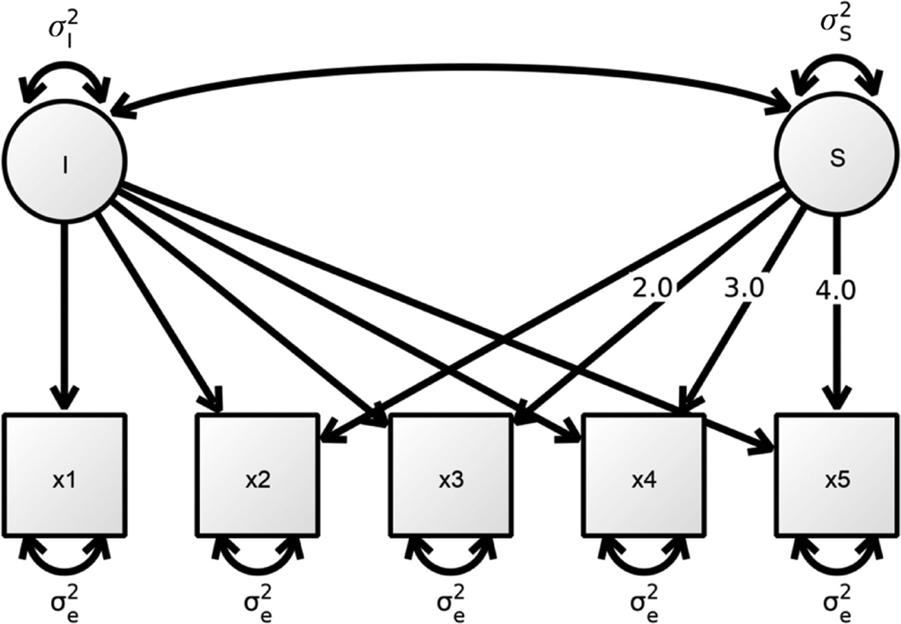
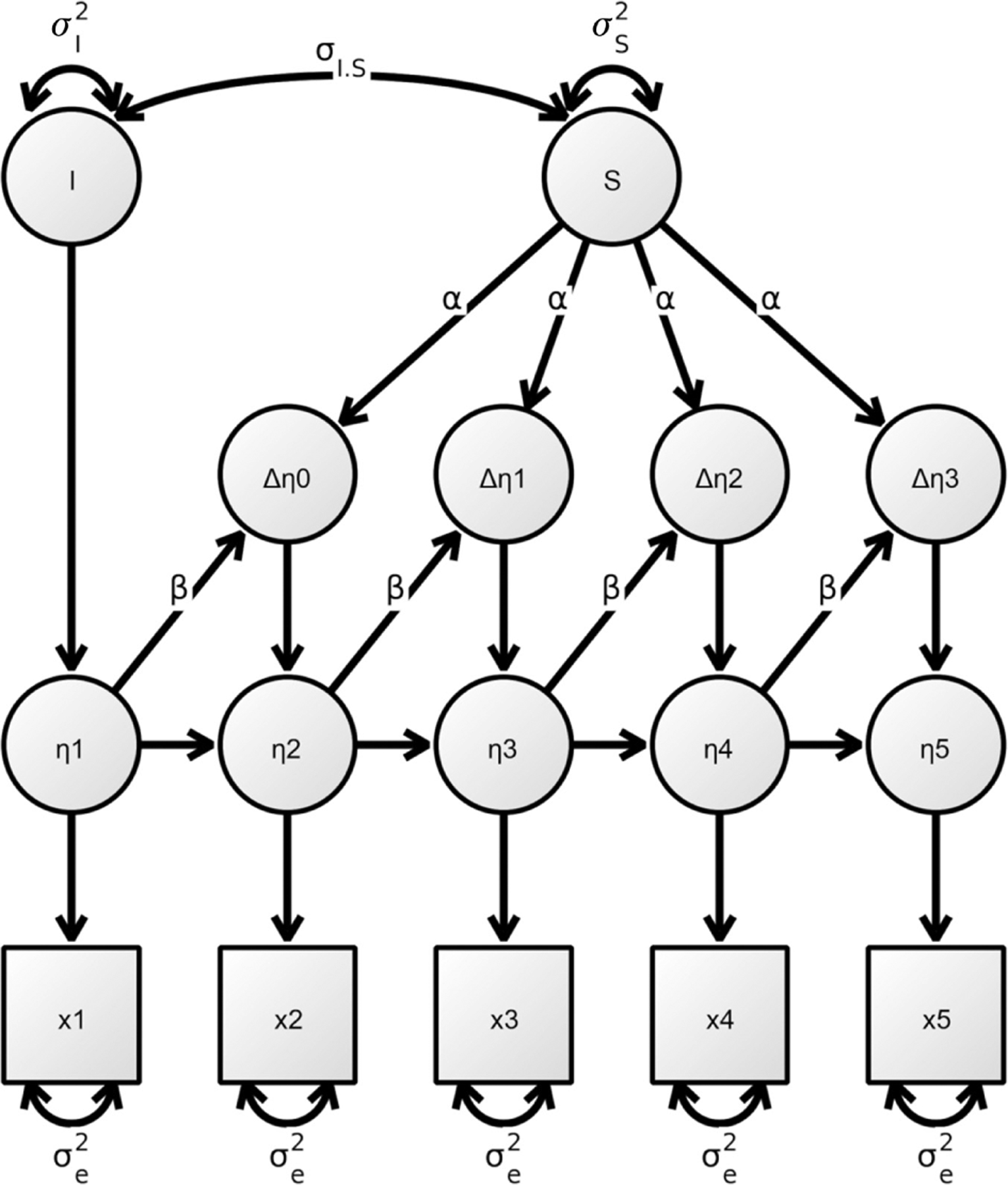
Little is still known about the neuroanatomical substrates related to changes in specific cognitive abilities in the course of healthy aging, and the existing evidence is predominantly based on cross-sectional studies. However, to understand the intricate dynamics between developmental changes in brain structure and changes in cognitive ability, longitudinal studies are needed. In the present article, we review the current longitudinal evidence on correlated changes between magnetic resonance imaging-derived measures of brain structure (e.g. gray matter/white matter volume, cortical thickness), and laboratory-based measures of fluid cognitive ability (e.g. intelligence, memory, processing speed) in healthy older adults. To theoretically embed the discussion, we refer to the revised Scaffolding Theory of Aging and Cognition. We found 31 eligible articles, with sample sizes ranging from n = 25 to n = 731 (median n = 104), and participant age ranging from 19 to 103. Several of these studies report positive correlated changes for specific regions and specific cognitive abilities (e.g. between structures of the medial temporal lobe and episodic memory). However, the number of studies presenting converging evidence is small, and the large methodological variability between studies precludes general conclusions. Methodological and theoretical limitations are discussed. Clearly, more empirical evidence is needed to advance the field. Therefore, we provide guidance for future researchers by presenting ideas to stimulate theory and methods for development.

Keywords: brain structure; change; cognitive ability; correlated change; healthy aging; longitudinal.


Conflict of interest statement

Conflict of interest statement: All authors declare no conflicts of interest.

Figures

Figure 1:
Figure 1:
Four different research questions on the relation between brain structure and cognitive ability, as illustrated with Cattell’s (1988) data box. Panel A: research question type 1 refers to interindividual differences in a measure of brain structure or cognitive ability assessed at one measurement occasion. Panel B: research question type 2 refers to intraindividual changes in a measure of brain structure or cognitive ability assessed across several measurement occasions. Panel C: research question type 3 refers to the bivariate association between interindividual differences in a measure of brain structure and interindividual differences in a measure of cognitive ability assessed at one measurement occasion (correlation). Panel D: research question type 4 refers to the bivariate association between intraindividual change in a measure of brain structure and intraindividual change in a measure of cognitive ability assessed across several measurement occasions (correlated change).
Figure 2:
Figure 2:
Scaffolding Theory of Aging and Cognition (STAC-r) model adapted from Reuter-Lorenz and Park (2014). *Under brain structure we subsume both structural brain properties and rate of brain structure change.
Figure 3:
Figure 3:
Potential cross-sectional (research question 3: correlation) and longitudinal (research question 4: level-change, simultaneous, and lagged correlated change) relations between brain structure (= Brain) and cognitive ability (= Cog). T = time/measurement occasion. ΔTn+1−Tn represents developmental change between two measurement occasions. Square shapes represent observed measures of a domain at a specific measurement occasion.
Figure 4:
Figure 4:
Preferred reporting items for systematic reviews and meta-analyses flow-chart of the literature search procedure.
Figure 5:
Figure 5:
A univariate latent growth curve model. Circles represent latent variables while squares represent manifest variables. One headed arrows denote directed relationships and double headed arrows represent undirected relationships. Here, I is the latent intercept and S is the latent slope, each with their corresponding variances σI2 and σS2.
Figure 6:
Figure 6:
A univariate latent change score model. x1–x5 represent the observed variable measured at five time points, η1–η5 represent latent true scores, Δη0–Δη4 are the latent change scores and I and S define the latent intercept and slope α paths represent constant change and β paths represent proportional change from the variable measured at the previous time point.

References

    1. Adólfsdóttir S, Wollschlaeger D, Wehling E, and Lundervold AJ (2017). Inhibition and switching in healthy aging: a longitudinal study. J. Int. Neuropsychol. Soc 23, 90–97. - PubMed
    1. Alexander-Bloch A, Clasen L, Stockman M, Ronan L, Lalonde F, Giedd J, and Raznahan A (2016). Subtle in-scanner motion biases automated measurement of brain anatomy from in vivo MRI. Hum. Brain Mapp 37, 2385–2397. - PMC - PubMed
    1. Anblagan D, Valdés Hernández MC, Ritchie SJ, Aribisala BS, Royle NA, Hamilton IF, Cox SR, Gow AJ, Pattie A, Corley J, et al. (2018). Coupled changes in hippocampal structure and cognitive ability in later life. Brain Behav. 8, e00838. - PMC - PubMed
    1. Arshad M, Stanley JA, and Raz N (2016). Adult age differences in subcortical myelin content are consistent with protracted myelination and unrelated to diffusion tensor imaging indices. Neuroimage 143, 26–39. - PMC - PubMed
    1. Ashburner J and Friston KJ (2000). Voxel-based morphometry – the methods. Neuroimage 11, 805–821. - PubMed

Web references

    1. Lifebrain (n.d). Retrieved May 16, 2019, from http://lifebrain.uio.no/.
    1. GitHub (n.d). Retrieved May 16, 2019, from http://github.com/.
    1. Open Science Framework (n.d). Retrieved May 16, 2019, from http://osf.io/.
    1. OpenNeuro (n.d). Retrieved May 16, 2019, from http://openneuro.org/.
    1. Open Neuroimaging Laboratory (n.d). Retrieved May 16, 2019, from http://openneu.ro/.

Publication types