. 2019 Jun 13;15(6):e1007790.
doi: 10.1371/journal.ppat.1007790. eCollection 2019 Jun.

Therapeutic efficacy of favipiravir against Bourbon virus in mice

Affiliations

Therapeutic efficacy of favipiravir against Bourbon virus in mice

Traci L Bricker et al. PLoS Pathog. .

Abstract

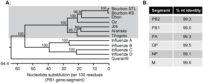
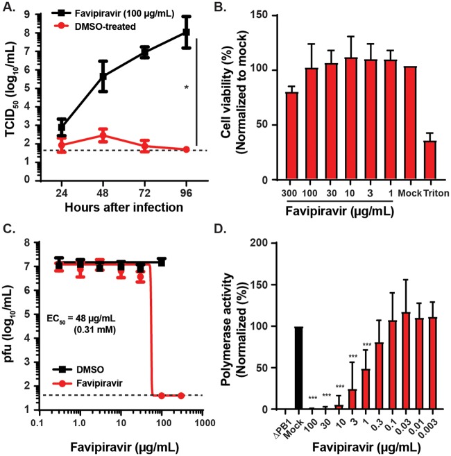
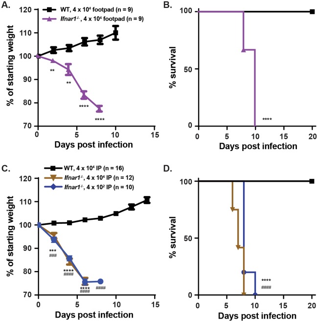
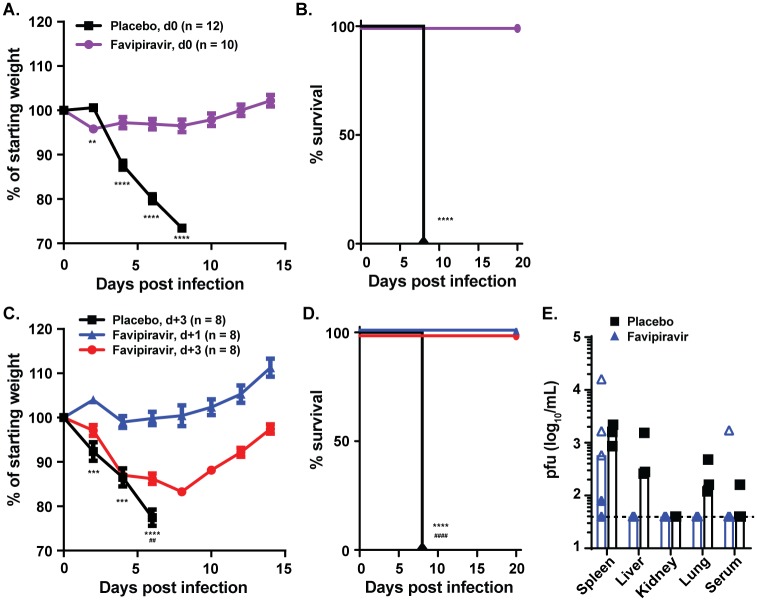
Bourbon virus (BRBV) is an emerging tick-borne RNA virus in the orthomyxoviridae family that was discovered in 2014. Although fatal human cases of BRBV have been described, little is known about its pathogenesis, and no antiviral therapies or vaccines exist. We obtained serum from a fatal case in 2017 and successfully recovered the second human infectious isolate of BRBV. Next-generation sequencing of the St. Louis isolate of BRBV (BRBV-STL) showed >99% nucleotide identity to the original reference isolate. Using BRBV-STL, we developed a small animal model to study BRBV-STL tropism in vivo and evaluated the prophylactic and therapeutic efficacy of the experimental antiviral drug favipiravir against BRBV-induced disease. Infection of Ifnar1-/- mice lacking the type I interferon receptor, but not congenic wild-type animals, resulted in uniformly fatal disease 6 to 10 days after infection. RNA in situ hybridization and viral yield assays demonstrated a broad tropism of BRBV-STL with highest levels detected in liver and spleen. In vitro replication and polymerase activity of BRBV-STL were inhibited by favipiravir. Moreover, administration of favipiravir as a prophylaxis or as post-exposure therapy three days after infection prevented BRBV-STL-induced mortality in immunocompromised Ifnar1-/- mice. These results suggest that favipiravir may be a candidate treatment for humans who become infected with BRBV.


Conflict of interest statement

The authors have declared that no competing interests exist.

Figures

Fig 1
Fig 1. Molecular characterization of BRBV-STL.
BRBV-STL was identified in a blood sample from a hospitalized patient. The genome sequence of the virus was determined by next-generation sequencing on RNA extracted from culture supernatant of Vero cells inoculated with the patient serum. (A) Phylogenetic tree of segment 2 (PB1 gene) of different orthomyxoviruses. The tree was constructed using ClustalW in the DNASTAR Lasergene 15 software package. Bootstrap values were calculated on a 1000 trails and the values are included in the figure. NA, no value available. Bourbon virus strain St. Louis (BRBV-STL, MK453528); Bourbon virus strain original (BRBV-KS, KU708254.1); Dhori virus (NC_034263.1); Oz virus (LC320124.1); Jos virus (HM627170.1); Aransas virus (KC506163.1); Thogoto virus (AF004985.1); Influenza A virus (CY009450); Influenza B virus (AY582058.1); Influenza C virus (NC_006308.2); Influenza D virus (LN559121.1); Quaranfil virus (FJ861695.1). BRBV-STL belongs to the Thogotovirus genus (grey background) and is very similar to the original strain of BRBV. (B) Percent identity at the nucleotide (nt) level between our BRBV isolate (BRBV-STL) and the original BRBV isolate (BRBV-KS).
Fig 2
Fig 2. Favipiravir inhibits BRBV-STL replication and polymerase activity.
(A) Vero cells were inoculated with 20 pfu of BRBV-STL in the presence or absence of 100 μg/mL (0.64 mM) favipiravir. The virus titer in the culture supernatant was quantified every 24 h for 4 days. Values are means (± standard deviation) of the virus titer from two experiments performed in duplicate. *, P < 0.05 by Mann-Whitney U-test on the slope of the curves fit by linear regression of ln-transformed virus titer over time. The dotted line represents the limit of detection at 50 TCID50/mL. (B) XTT assay to measure Vero cell viability at different doses of favipiravir or solvent (DMSO). The results are normalized to untreated (Mock) control cells. Triton X-100 (1%, Triton) was used as a positive control. Values are the means (+ standard deviation) of the normalized cell viability data. Each condition was tested in triplicate, and each experiment was repeated three independent times. (C) Vero cells were inoculated with 20 pfu of BRBV-STL in the presence of different concentrations of favipiravir or DMSO (solvent), starting at 300 μg/mL (1.9 mM). The virus titer in the culture supernatant was quantified by plaque assay at 3 dpi. Values are geometric means (± standard deviation) of the virus titer from three experiments performed in duplicate. A curve was fitted through the data using the log(inhibitor) vs. response—variable slope equation (GraphPad Prism 8.0). The dotted line represents the limit of detection at 40 pfu/mL. (D) The effect of different concentrations of favipiravir on the polymerase activity was quantified in a mini-genome reporter assay. Expression plasmids encoding for the PB2, PB1, PA and NP of BRBV-STL and Renilla luciferase, plus the reporter construct (Firefly luciferase flanked by the 3' and 5' UTR of segment 5 of BRBV-STL) were transfected into 293T cells in the presence or absence of different concentrations of favipiravir. Three days post transfection the amount of firefly luciferase was quantified and normalized across wells and conditions using the Renilla luciferase data. Values are the means (+ standard deviation) of the normalized polymerase activity from seven experiments performed in duplicate. ***, P < 0.001 by one-way ANOVA correcting for multiple comparisons.
Fig 3
Fig 3. Mouse model of BRBV disease.
(A and B) WT (n = 9), and Ifnar1-/- mice (n = 9), were inoculated with 4 x 10⁴ pfu of BRBV-STL in the footpad. Weight change (A) and survival (B) were monitored for 10 and 20 days respectively. Values are the means (± standard error of the mean) from two experiments. Weight change is analyzed by an independent t-test and survival by the log-rank test. **, P < 0.01; ****, P < 0.0001. (C and D) WT and Ifnar1-/- mice were inoculated via intraperitoneal route with 4 x 10⁴ (n = 16 for WT and n = 12 for Ifnar1-/-) or 4 x 10² (n = 10 for Ifnar1-/-) pfu of BRBV-STL and weight change (C) and survival (D) were monitored for 14 and 20 days respectively. Values are the means (± standard error of the mean) from two or more experiments. Weight change is analyzed by an independent t-test and survival by the log-rank test. ***, P < 0.001, ****, P < 0.0001 between WT and Ifnar1-/- mice at 4 x 10⁴ pfu. ###, P < 0.001; ####, P < 0.0001 between WT at 4 x 10⁴ pfu and Ifnar1-/- mice at 4 x 10² pfu.
Fig 4
Fig 4. Liver and spleen tropism of BRBV-STL in Ifnar1-/- mice.
(A and B) Virus titers (pfu/mL) were quantified in the liver, spleen, kidney, lung and serum of WT and Ifnar1-/- mice 3 and 6 dpi after IP inoculation with 4 x 10⁴ pfu of BRBV-STL. Tissues were collected, homogenized, and the virus titer was determined by plaque assay. Each data point is a single mouse obtained from two different experiments (indicated by the open and closed symbols). The bar represents the median virus titer observed in each of the tissues. Viremia in serum at 3 dpi was obtained from two mice in one experiment. The dotted line represents the limit of detection of the assay at 40 pfu/mL. (C) RNA in situ hybridization on sections from liver and spleen from uninfected of BRBV-STL infected (4 x 10⁴ pfu of BRBV-STL via IP) Ifnar1-/- mice. Probes targeting segment 5 of BRBV were used to visualize BRBV-infected cells, indicated by the dark brown staining. Sections were counterstained with hematoxylin prior to mounting and analysis. From left to right are uninfected, 3 dpi and 6 dpi at 10x and 40x magnification. Top panel are liver sections and the bottom panel are spleen sections. The images are representative of sections obtained from three mice in one experiment.
Fig 5
Fig 5. Favipiravir protects mice from fatal BRBV infection.
(A and B) Ifnar1-/- mice were treated with 150 mg/kg of favipiravir in 0.5% methylcellulose twice daily per oral gavage (n = 10) or 0.5% methylcellulose alone (n = 12) for eight days starting 2 hours prior to infection with 4 x 10² pfu of BRBV-STL via IP. Weight change (A) and survival (B) were monitored for 14 and 20 days respectively. Values are the means (± standard error of the mean) from three experiments. Weight change is analyzed by an independent t-test and survival by the log-rank test. **, P < 0.01; ****, P < 0.0001. (C and D) Ifnar1-/- mice were treated eight days with 150 mg/kg of favipiravir in 0.5% methylcellulose twice daily starting one day (d+1, blue symbols, n = 8) or three days (d+3, red symbols, n = 8) after IP inoculation with 4 x 10² pfu of BRBV-STL. Mice receiving 0.5% methylcellulose alone for eight days starting three days after inoculation (placebo d+3, n = 8) were used as controls. Weight change (C) and survival (D) were monitored for 14 and 20 days respectively. Values are the means (± standard error of the mean) from two experiments. Weight change is analyzed by an independent T-test and survival by the log-rank test. ***, P < 0.001; ****, P < 0.0001 between placebo and d+1 treated animals; ##, P < 0.01, ####, P < 0.0001 between placebo and d+3 treated animals. (E) Virus titers (pfu/mL) were quantified in various organs of mice infected with 4 x 10² pfu of BRBV-STL and treated one day after infection with 150 mg/kg of favipiravir twice daily for three days (n = 5) or 0.5% methylcellulose alone (n = 3). Each data point is a single mouse obtained from two different experiments (indicated by the open and closed symbols). The bar represents the median virus titer observed in each of the tissues. The dotted line represents the limit of detection of the assay at 40 pfu/mL.

Similar articles

Cited by

References

    1. Kosoy OI, Lambert AJ, Hawkinson DJ, Pastula DM, Goldsmith CS, Hunt DC, et al. Novel thogotovirus associated with febrile illness and death, United States, 2014. Emerg Infect Dis. 2015;21(5):760–4. 10.3201/eid2105.150150 - DOI - PMC - PubMed
    1. Savage HM, Burkhalter KL, Godsey MS Jr., Panella NA, Ashley DC, Nicholson WL, et al. Bourbon Virus in Field-Collected Ticks, Missouri, USA. Emerg Infect Dis. 2017;23(12):2017–22. Epub 2017/11/18. 10.3201/eid2312.170532 . - DOI - PMC - PubMed
    1. Lambert AJ, Velez JO, Brault AC, Calvert AE, Bell-Sakyi L, Bosco-Lauth AM, et al. Molecular, serological and in vitro culture-based characterization of Bourbon virus, a newly described human pathogen of the genus Thogotovirus. J Clin Virol. 2015;73:127–32. 10.1016/j.jcv.2015.10.021 Epub Oct 27. - DOI - PMC - PubMed
    1. Savage HM, Godsey MS Jr., Panella NA, Burkhalter KL, Manford J, Trevino-Garrison IC, et al. Surveillance for Tick-Borne Viruses Near the Location of a Fatal Human Case of Bourbon Virus (Family Orthomyxoviridae: Genus Thogotovirus) in Eastern Kansas, 2015. Journal of medical entomology. 2018;55(3):701–5. Epub 2018/01/25. 10.1093/jme/tjx251 . - DOI - PubMed
    1. Furuta Y, Takahashi K, Kuno-Maekawa M, Sangawa H, Uehara S, Kozaki K, et al. Mechanism of action of T-705 against influenza virus. Antimicrob Agents Chemother. 2005;49(3):981–6. Epub 2005/02/25. 10.1128/AAC.49.3.981-986.2005 . - DOI - PMC - PubMed

Publication types

MeSH terms